
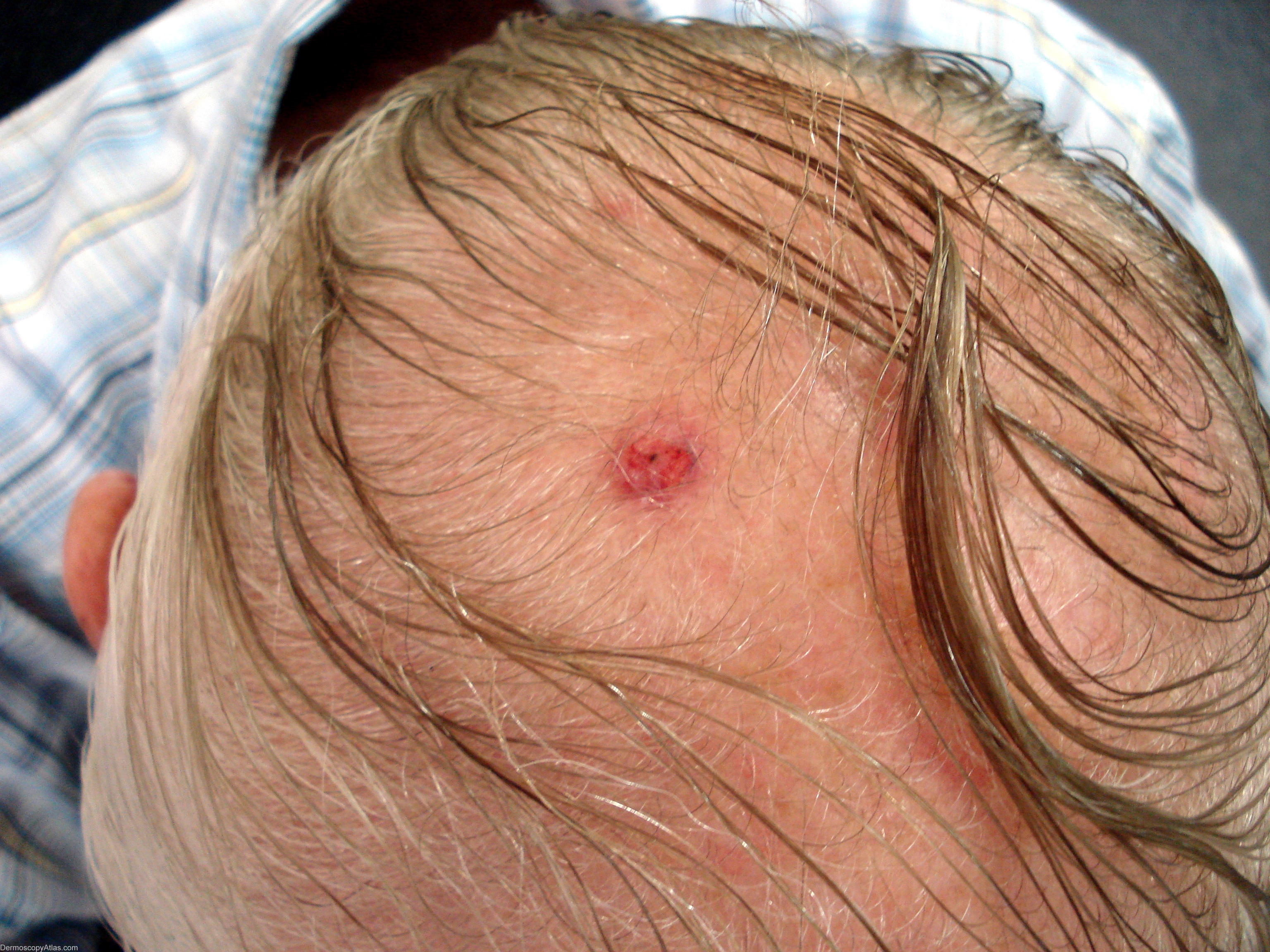
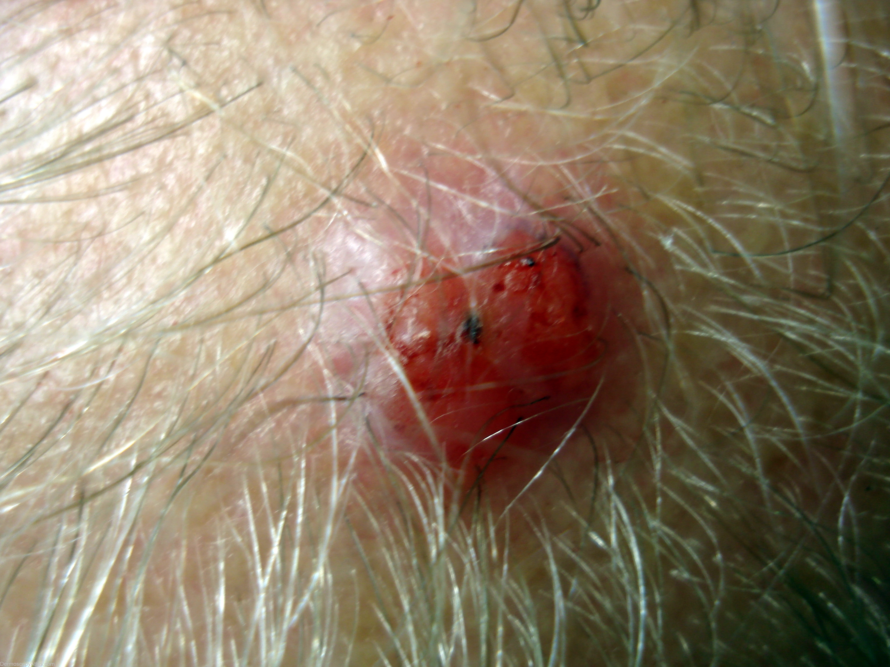
Site: Scalp
Diagnosis: Atypical fibroxanthoma
Sex: M
Age: 76
Type: Heine
Submitted By:
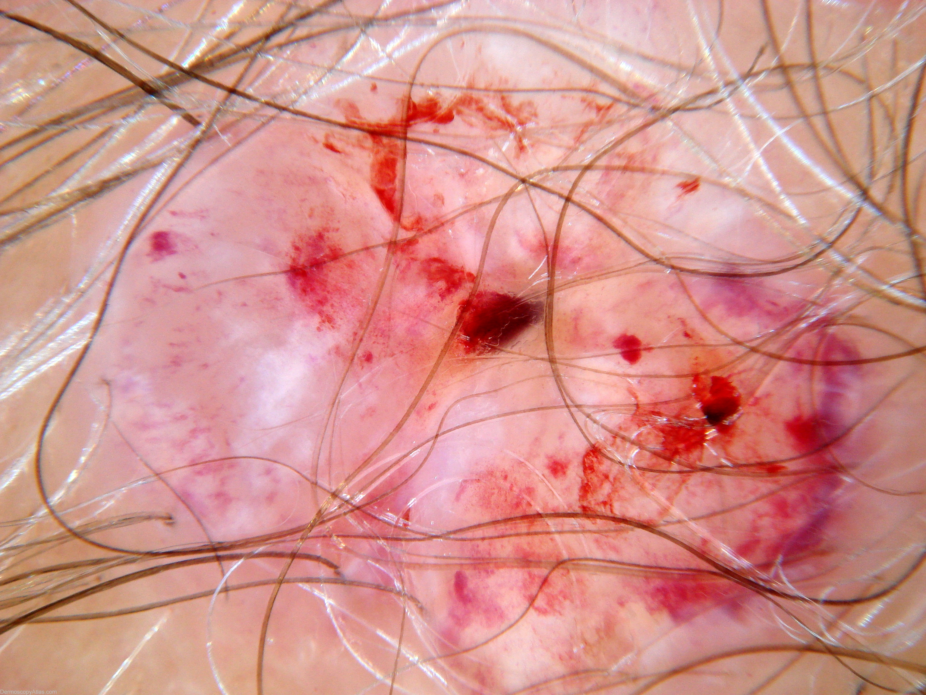
Description: Dermoscopy view - (Editor's comment - CR). This is an intradermal,spindle cell tumour. It exhibits low to moderate malignant potential and recurrences are most commonly due to incomplete excision. Regional metastases, though rare, tend to occurr in large ulcerated lesions, in previously irradiated sites or in immunocompromised patients.( Ref Cancer of the Skin - Rigel page 541). This lesion shows white areas (much whiter than surrounding skin), significant ulceration, no apparent pigmentation but a variety of blood vessel types including hairpin vessels. dot vessels and linear vessels.
History: 76 yr old man with slowly enlarging lesion on scalp. Excised and repaired with FTSG