
Site: Back
Diagnosis: BCC pigmented
Sex: F
Age: 62
Type: Dermlite Non Polarised
Submitted By: Cliff Rosendahl
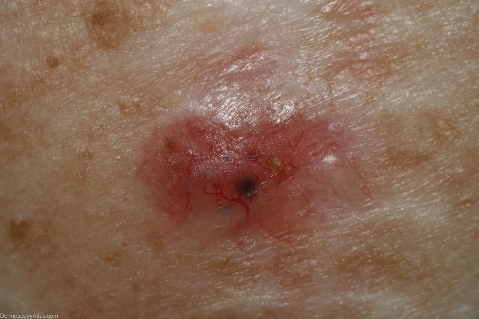
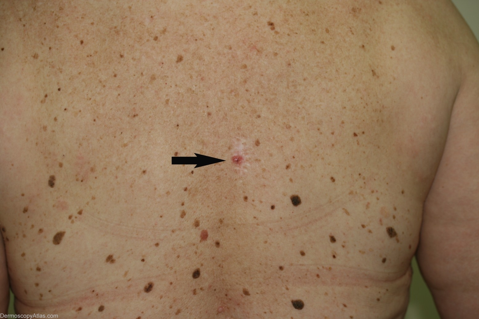
Description: Clinical image - This image shows this BCC to be arising in continuity with a surgical scar. That scar was from a BCC excision 5 years earlier which was reported as "clear of excision margins"
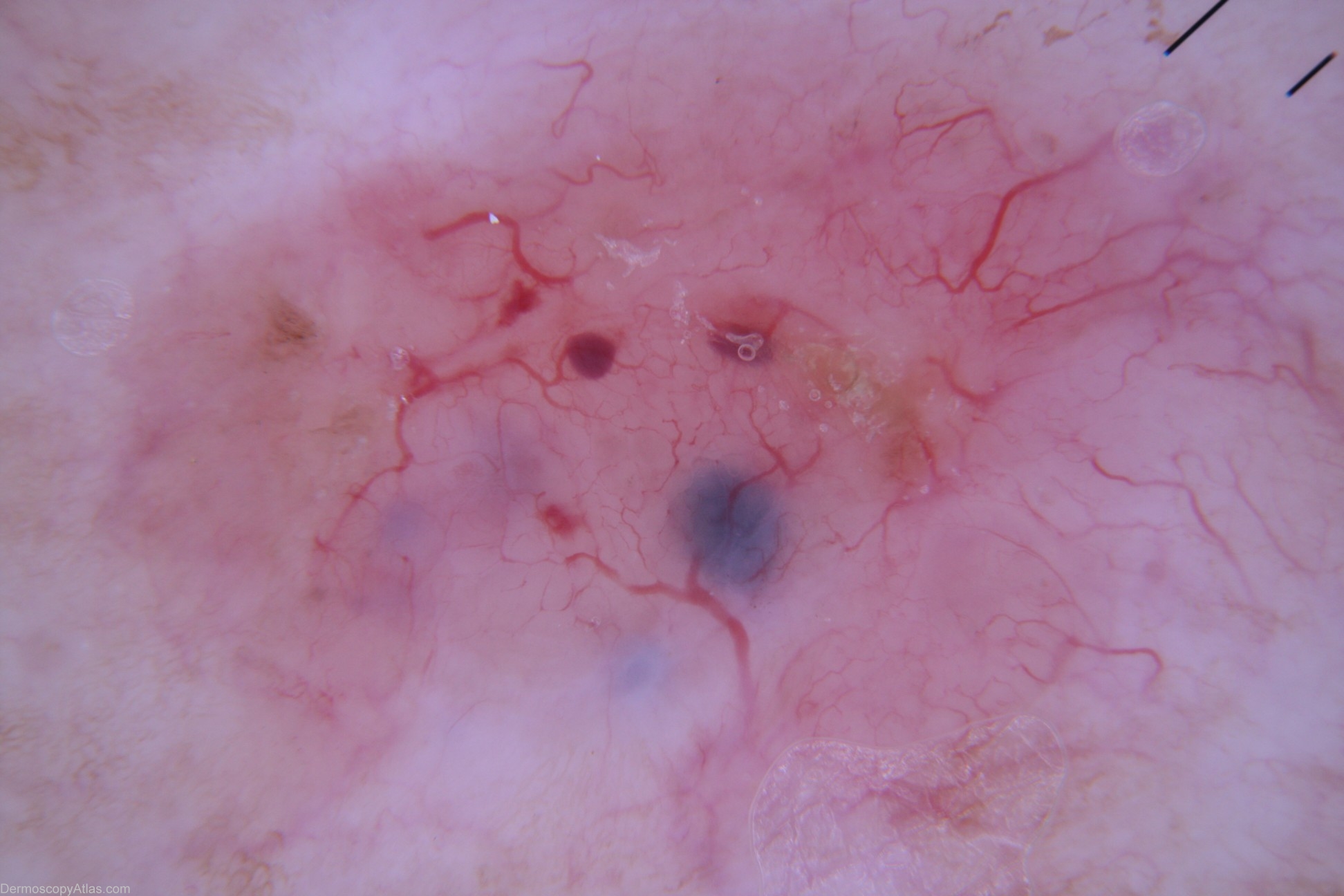
History: This lesion was encountered at a routine skin check on this new patient to the practice. Histology was reported as "...a focally pigmented BCC of solid type expanding the papillary dermis". It was excised with 10mm margins as it was apparently a recurrent BCC arising in continuity with a surgical scar from a BCC excision 5 years earlier.