
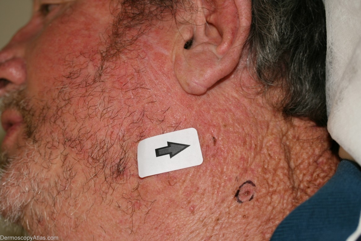
Site: Neck side
Diagnosis: Lentigo Maligna
Sex: M
Age: 62
Type: Dermlite Non Polarised
Submitted By: Cliff Rosendahl
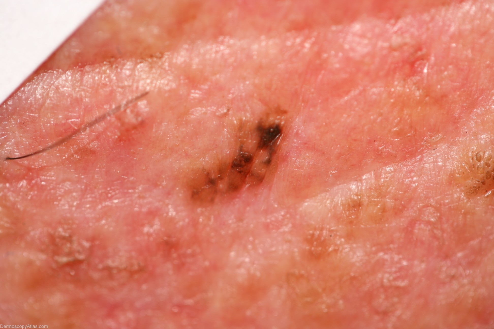
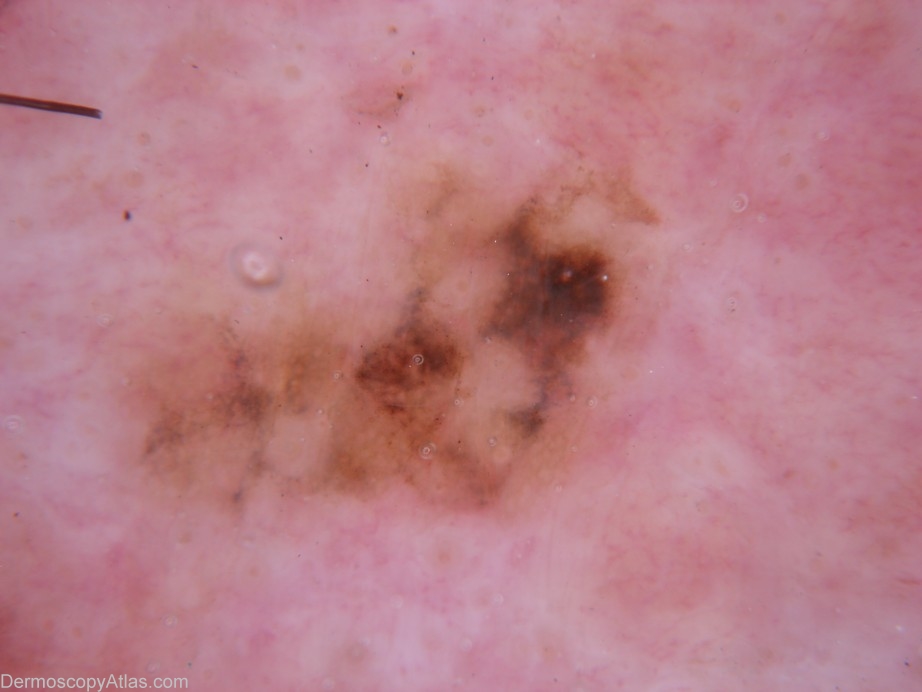
Description: Enter Description This Lentigo maligna ehhibits asymmetry and the presence of more than one colour. There are blue-grey dots (peppering), a focus of broadened pigment network and several structureless areas interspersed with patches of reticular network
History: This 62 year old man had attended 3 weeks earlier for a skin check and I had identified a suspected BCC and scheduled a biopsy. He attended for that biopsy and this pigmented lesion was noted. I had not seen it 3 weeks earlier and I suspect it must have become much more evident over that period of 3 weeks. Shave biopsy was reported as lentigo maligna and the lesion was subsequently re-excised with greater than 5 mm margins.