
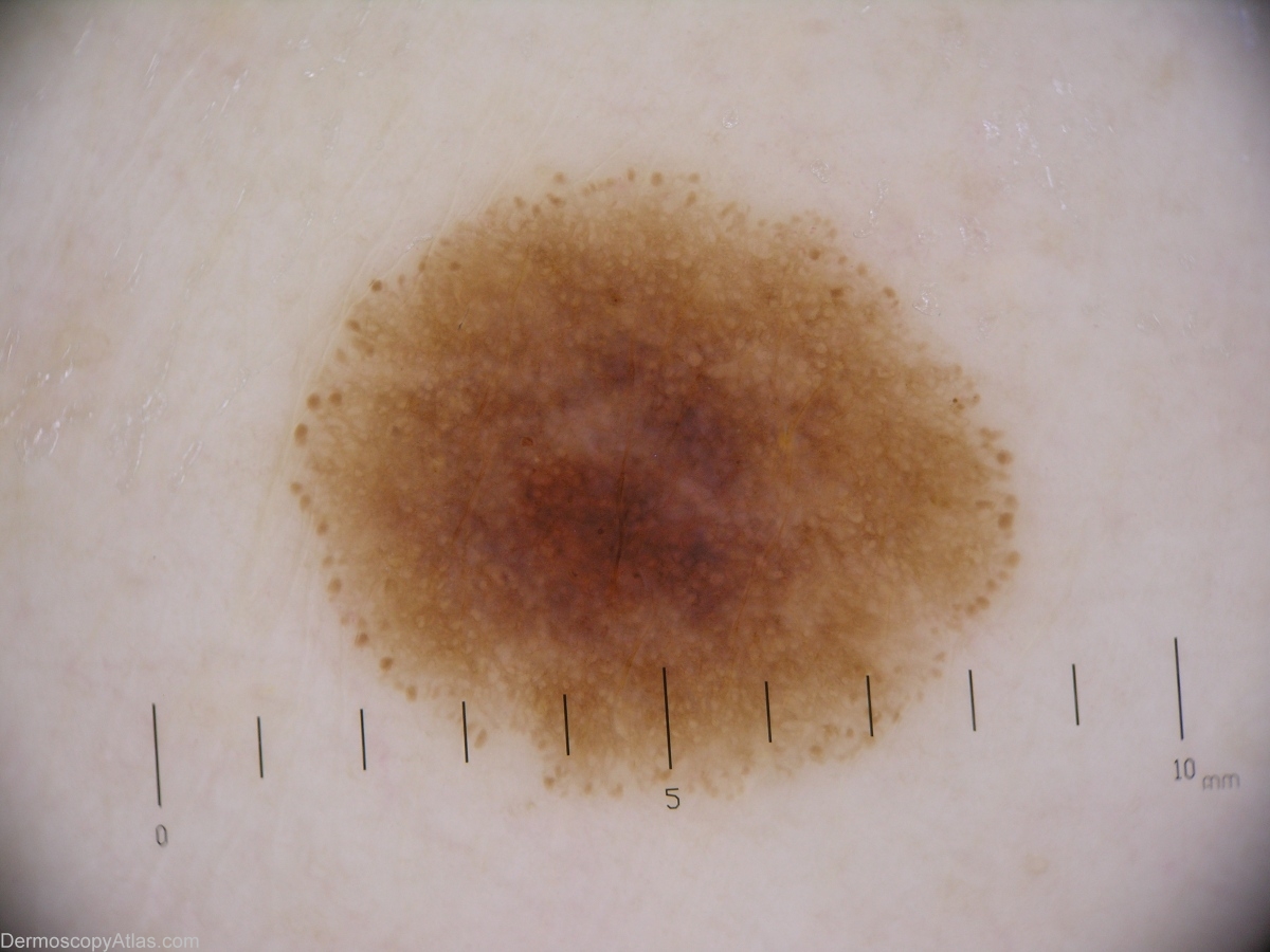
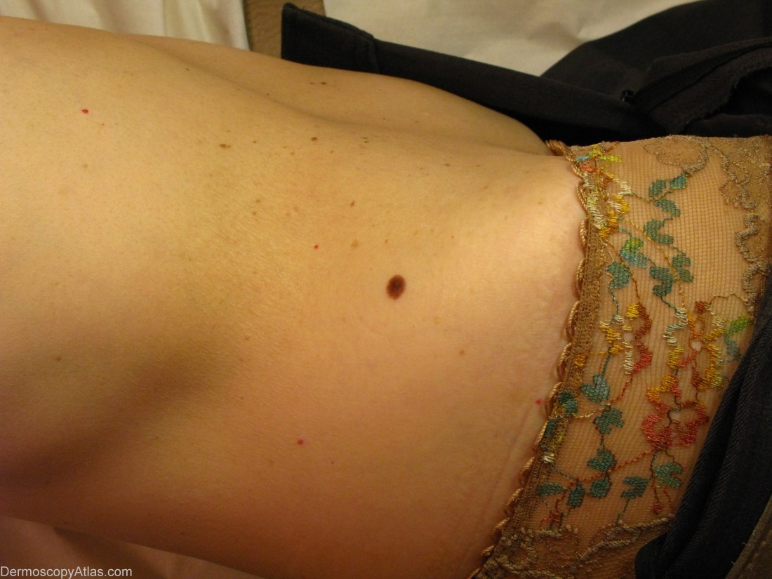
Site: Abdomen
Diagnosis: Nevus compound
Sex: F
Age: 60
Type: Heine
Submitted By: Jean-Yves Gourhant
Description: A regular network, surrounded by a symmetrically disposed rim of brown globules.
History:
This 60 years old lady came for this mole, which was located just above her right hip. She had noticed it only recently, less than 6 months, and thought it was enlarging.
The dermoscopic exam showed a regular network, which was slightly darker in the center, and all around a rim of regularly distributed brown globules. This aspect was strongly in favour of a benign enlarging nevus.
Owing to her age, the lesion was excised, and the pathology confirmed the clinical diagnosis of compound nevus
Enlarging common melanocytic nevus are relatively uncommon in older age. In the study of Harald Kittler, it is said that nearly half of these nevus show histological signs of atypia, which was not the case here.Reference: "Frequency and Characteristics of Enlarging Common Melanocytic nevi", Kittler and al, Archives of Derm; vol 136, march 2000:316-320.