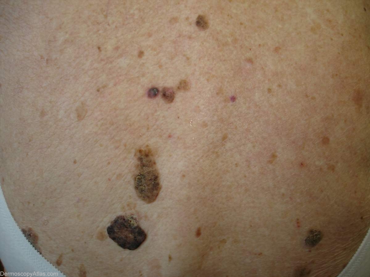
Loading
TeleDermConsult-logo
  • Recent Submissions
  • Search
  • Diagnosis
  • Quiz
  • Image comparison
  • Submit
  • Teaching Cases
  • Index
  • About

<< Back to Diagnosis Page

Image Number #2609 (Pigmented basal cell carcinoma)

Site: Back

Diagnosis: Pigmented basal cell carcinoma

Sex: F

Age: 75

Type: Dermlite Polarised

Submitted By: Ian McColl

View Full Size Image

Click Here to View Details

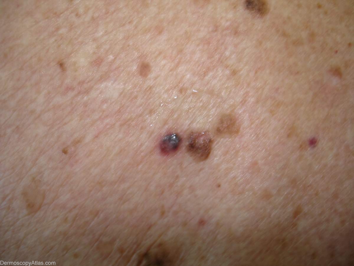
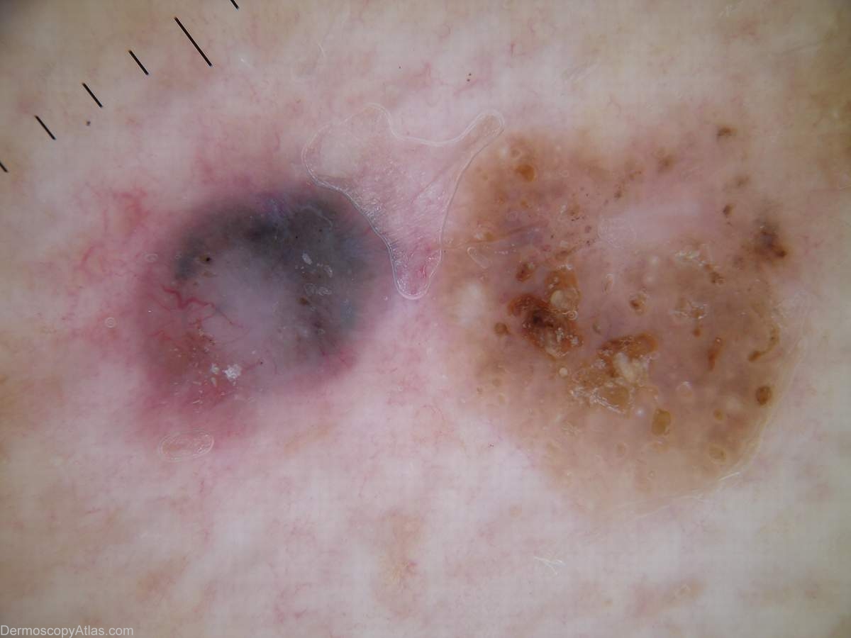
Description: Comparison of two pigmented lesions Note the blue grey ovoid nests and the arborising vessels of the pigmented BCC and the crypts and milia of the seborrhoeic keratosis. Harald would describe these as orange and white clods.

History: This lady had multiple seborrhoeic keratoses on her back but sometimes a lesion sticks out as a bit unusual. This was the juxtaposition of a deeply pigmented BCC and a seborrhoeic keratosis.

DermNetNZ   eMedicine   PubMed   Dermatology Online   Archives   JAAD for "Pigmented basal cell carcinoma"

  • Recent Submissions
  • Search
  • Diagnosis
  • Quiz
  • Image comparison
  • Submit an Image
  • Teaching Cases
  • Index
  • About

Copyright 2022. Dermoscopy Atlas. ALL Rights Reserved.

Change Image