
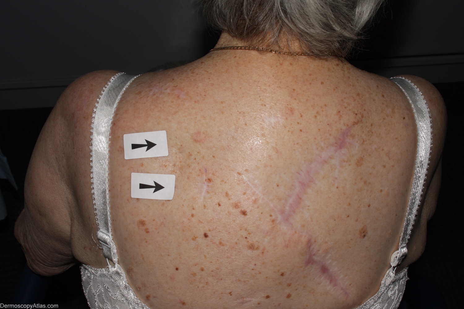
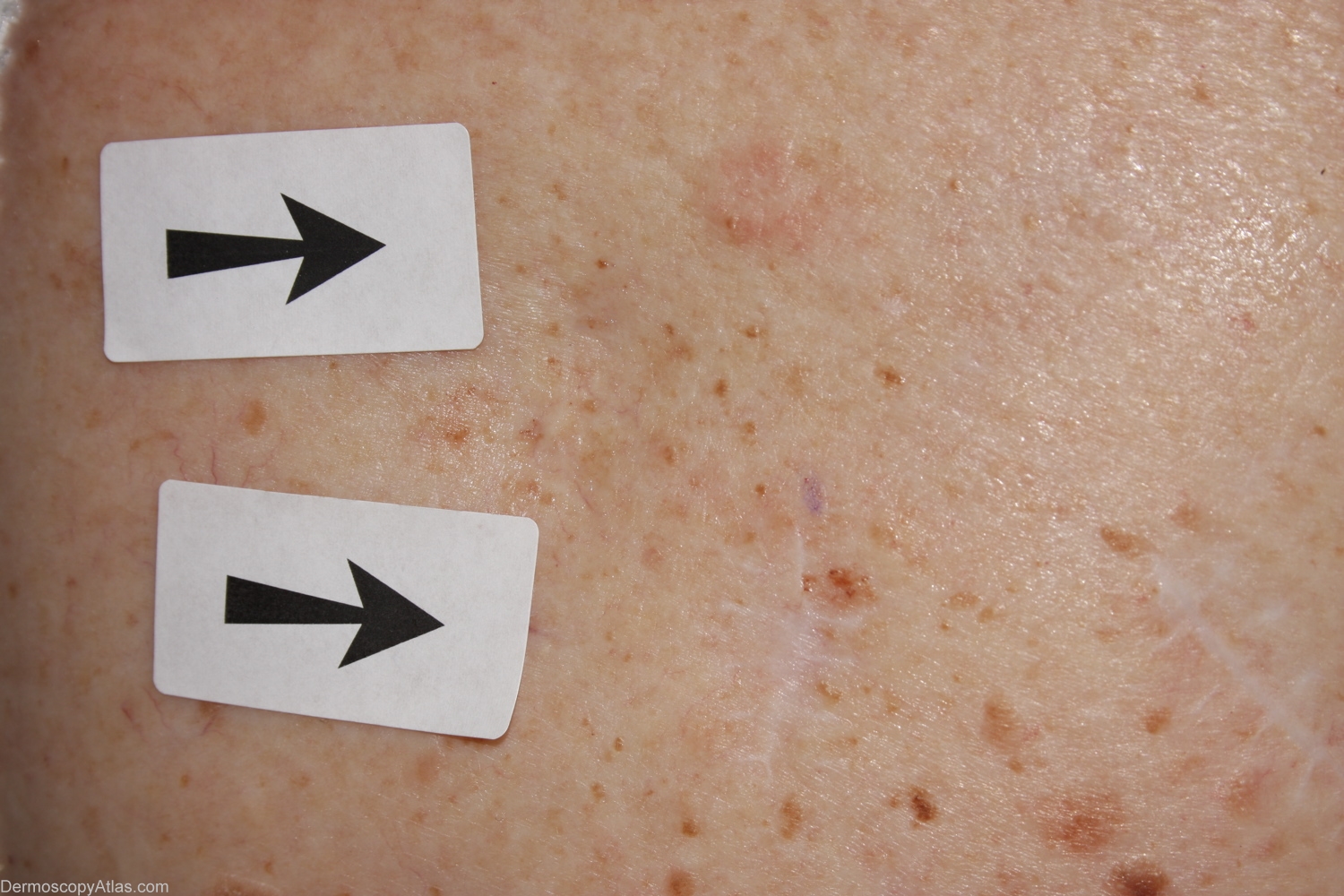
Site: Back
Diagnosis: Melanoma amelanotic
Sex: F
Age: 79
Type: Mixed
Submitted By: Alan Cameron
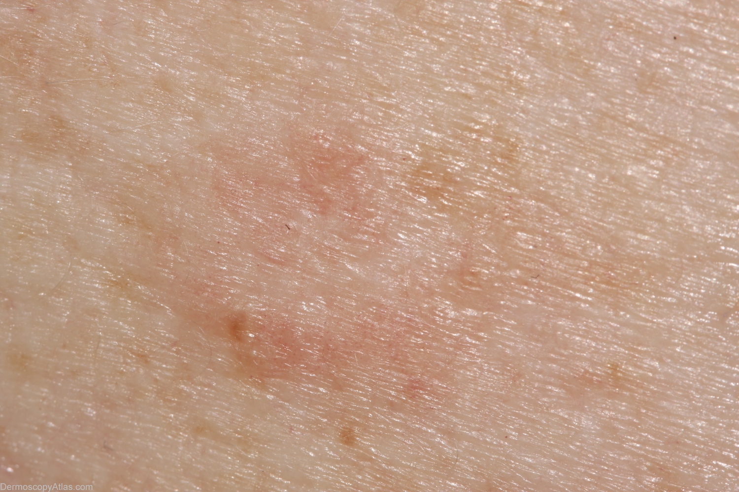
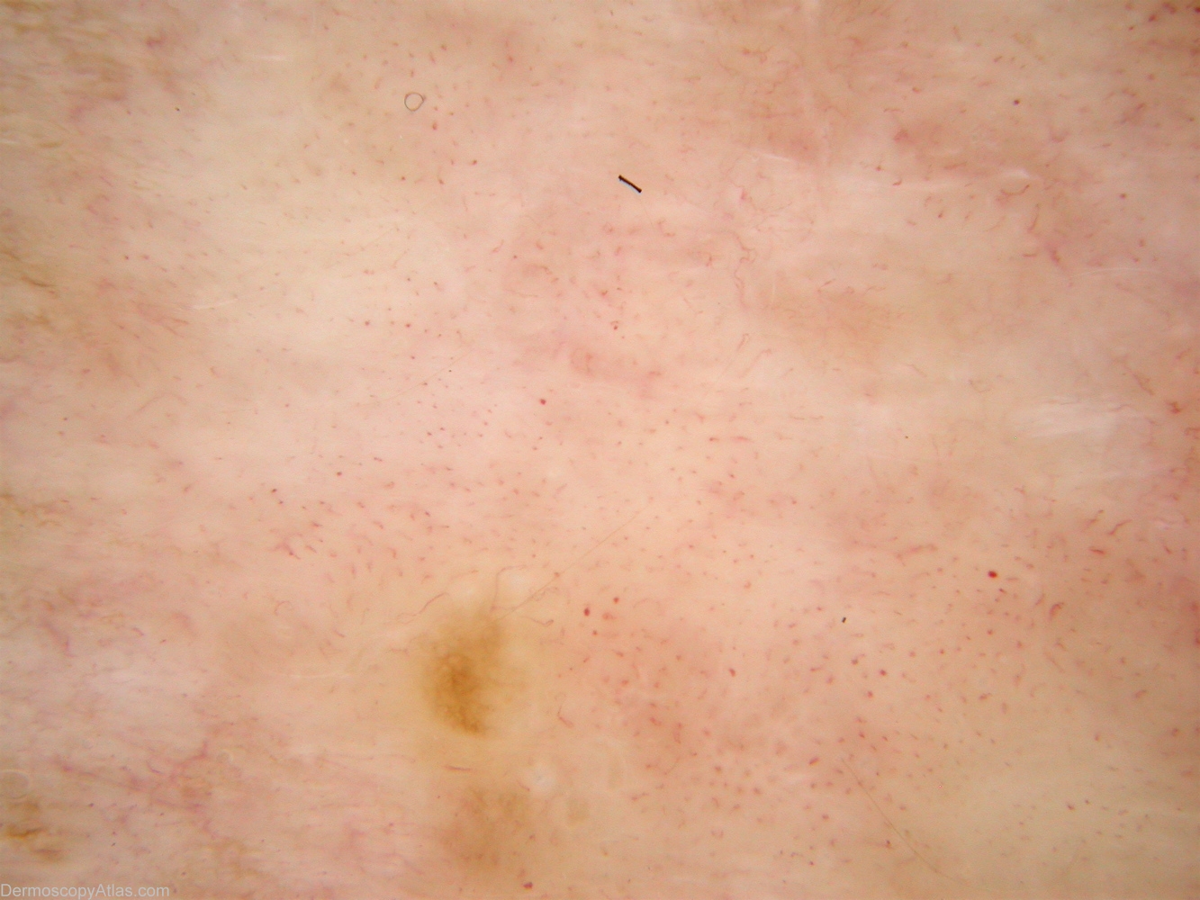
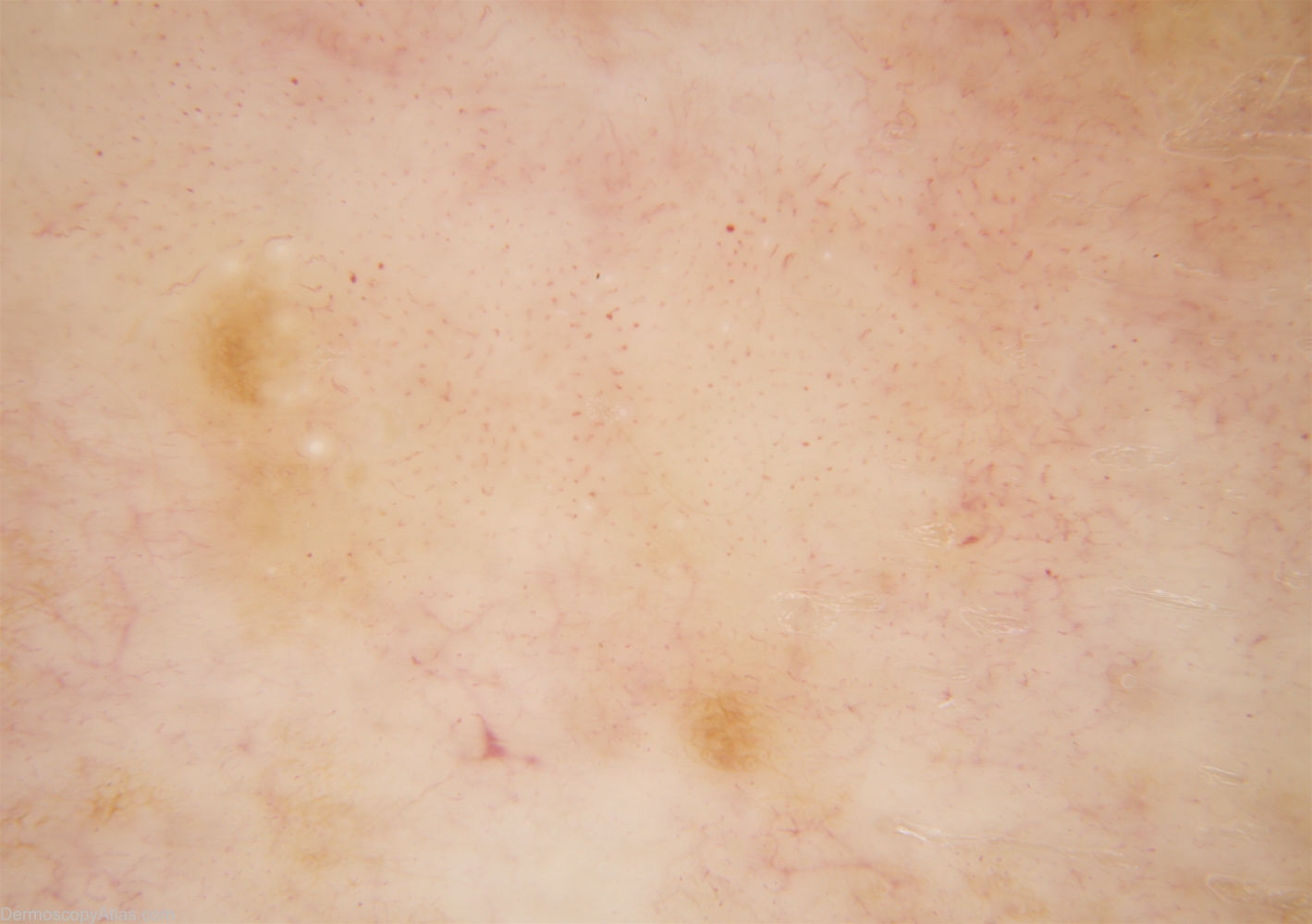
Description: Non-polarised Dermlite Fluid. Polymorphous vessels are better displayed on polarising dermoscopy. A few milia cysts (not seen on polarised dermoscopy) at 10 o'clock are unusual in melanoma but are sometimes found in BCC. Image enhancement to better display vessels has greatly exaggerated lesions pigmentation
History: This 79 year old woman has had 9 previous melanomas. Two lesions marked for biopsy, lesion in question superior. Histology was reported as — Sections show a few nests of superficial basal cell carcinoma admixed with a very focal level 2 (0.3mm thick) lentigo maligna melanoma. It is arising in a larger in situ lesion. There is no ulceration or dermal mitoses. There is a mild lymphocytic infiltrate but little regression.