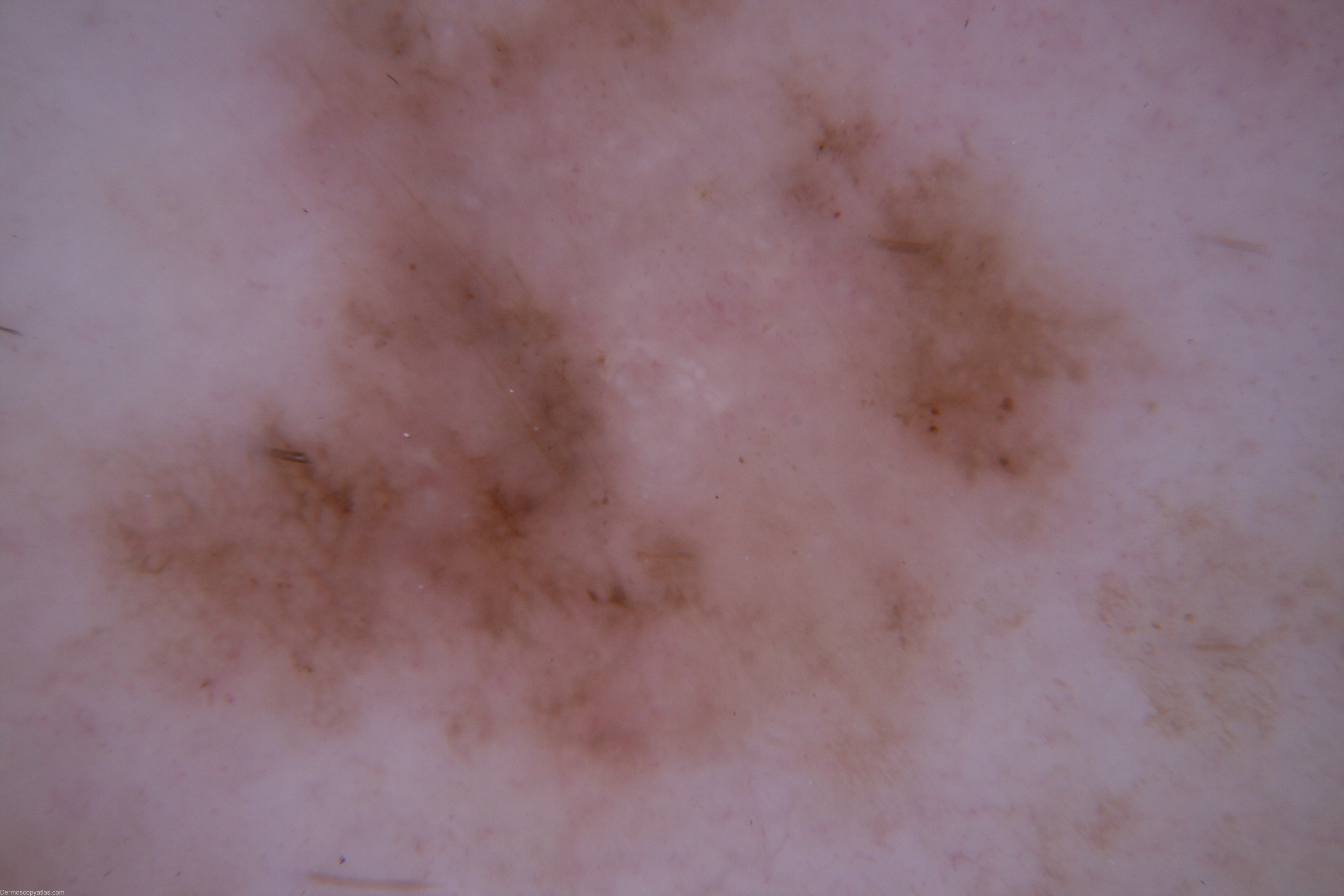
Loading
TeleDermConsult-logo
  • Recent Submissions
  • Search
  • Diagnosis
  • Quiz
  • Image comparison
  • Submit
  • Teaching Cases
  • Index
  • About

<< Back to Diagnosis Page

Image Number #2556 (Melanoma invasive)

Site: Leg

Diagnosis: Melanoma invasive

Sex: M

Age: 45

Type: Dermlite Non Polarised

Submitted By: Cliff Rosendahl

View Full Size Image

Click Here to View Details

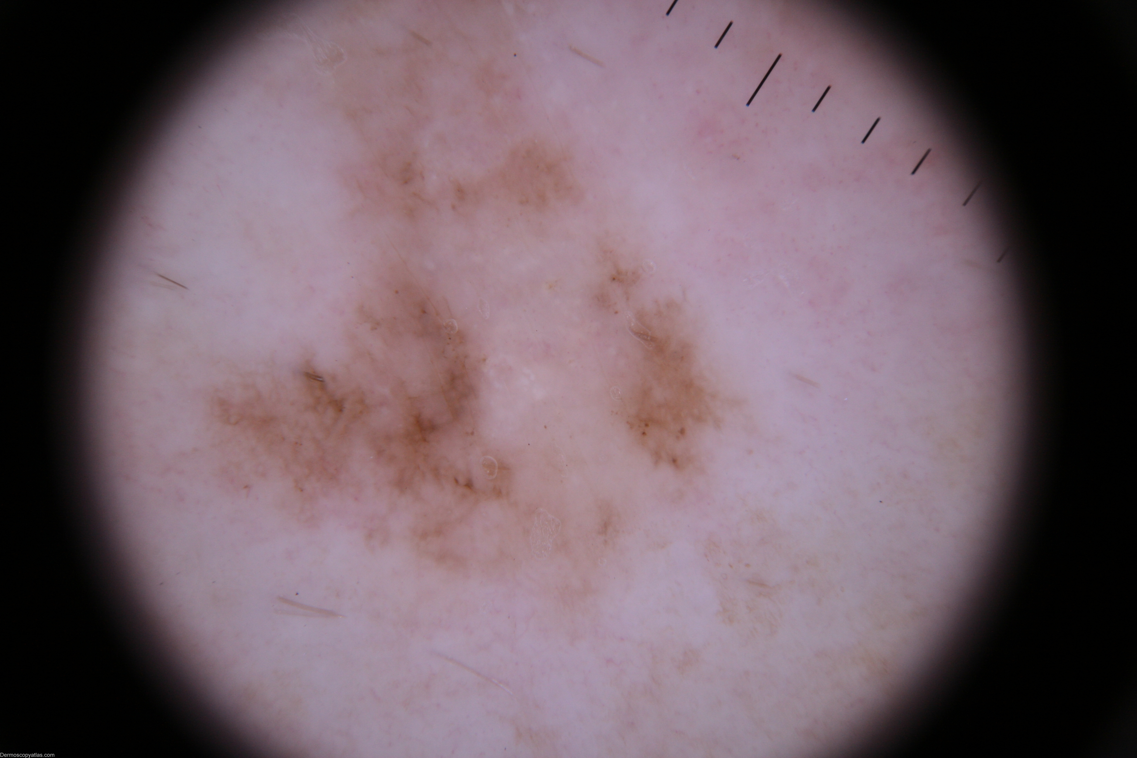
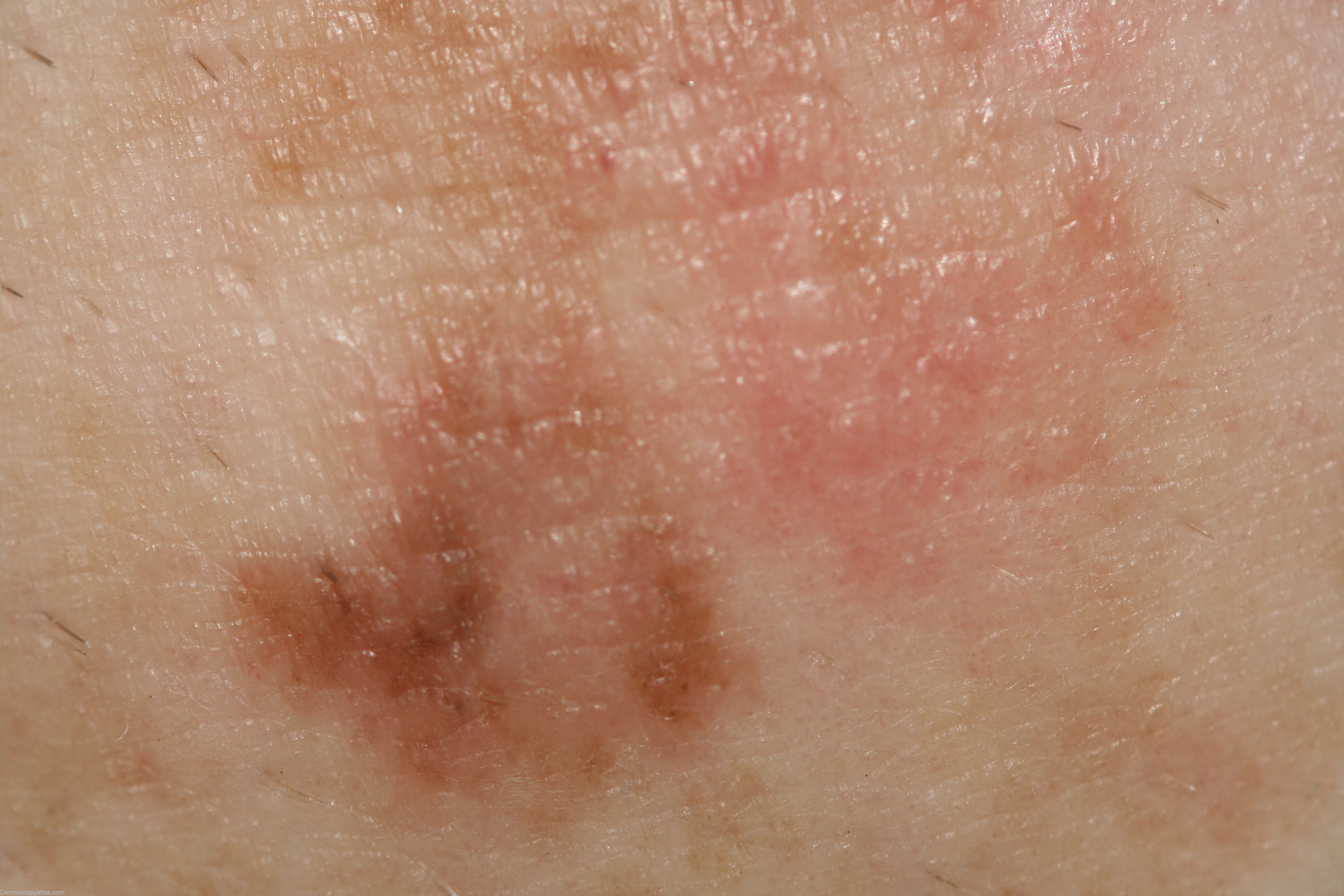
Description: > 1 pattern: lines reticular, structureless zone, >1 color clue to melanoma: structureless white zone, eccentric

History: This 45 year old man with a past history of multiple BCC,s presented for a skin examination 18 months after his previous examination. Histologically this was reported as "...level 2 (Breslow thickness 0.49mm) superficial spreading melanoma and solid BCC..."

DermNetNZ   eMedicine   PubMed   Dermatology Online   Archives   JAAD for "Melanoma invasive"

  • Recent Submissions
  • Search
  • Diagnosis
  • Quiz
  • Image comparison
  • Submit an Image
  • Teaching Cases
  • Index
  • About

Copyright 2022. Dermoscopy Atlas. ALL Rights Reserved.

Change Image