
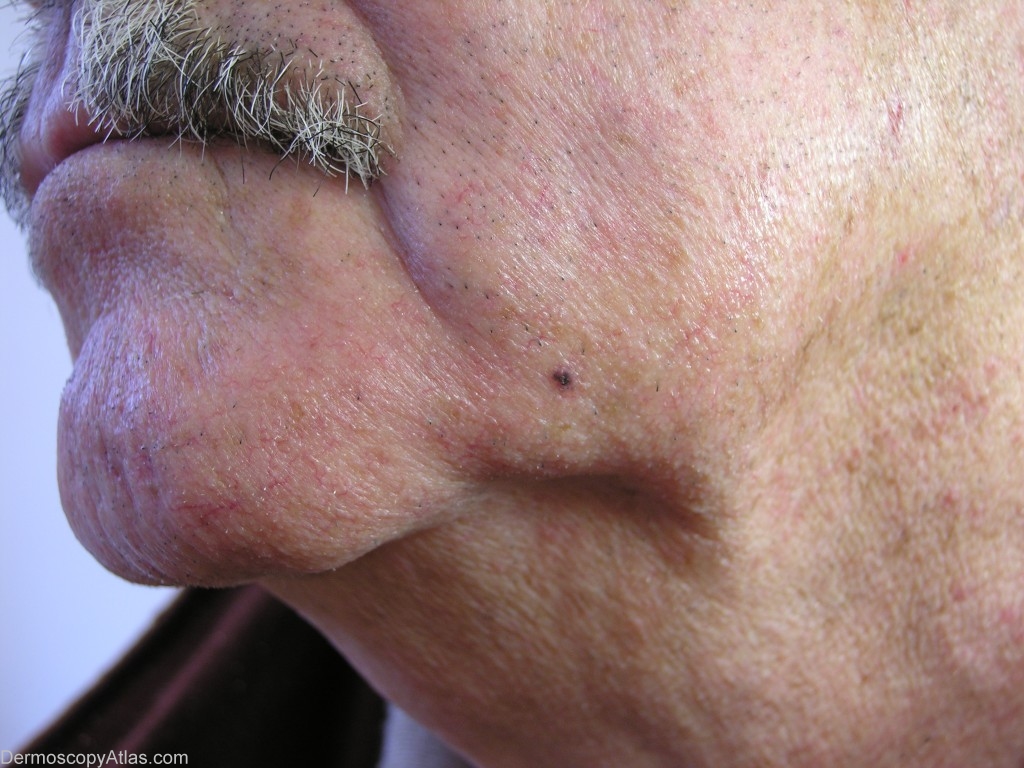
Site: Chin
Diagnosis: Pigmented basal cell carcinoma
Sex: M
Age: 81
Type: Heine
Submitted By: Jean-Yves Gourhant
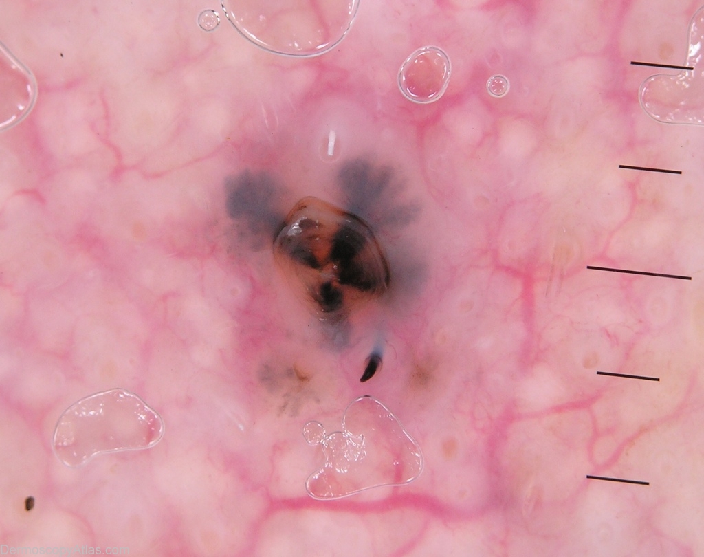
Description: The leaf-like area (here rather clover shape than maple leaf). Ulceration in the centre. The big vessels surrounding the lesion correspond to erythrosis.
History: This small pigmented, ulcerated lesion was observed during the exam of this 81 years old gentleman. The pathology confirmed the clinical diagnosis of nodular basal cell carcinoma. The dermoscopy shows a single small pigmented leaf-like area and an ulceration. Concerning the diagnosis of basal cell carcinoma,the specificity of the pigmented leaf-like area is 100% (Menzies et al, Surface microscopy of pigmented basal cell carcinoma.Arch Dermatol 2000: 1012-16).