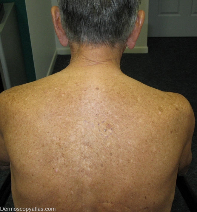
Loading
TeleDermConsult-logo
  • Recent Submissions
  • Search
  • Diagnosis
  • Quiz
  • Image comparison
  • Submit
  • Teaching Cases
  • Index
  • About

<< Back to Diagnosis Page

Image Number #2503 (BCC pigmented)

Site: Back

Diagnosis: BCC pigmented

Sex: M

Age: 81

Type: Dermlite Non Polarised

Submitted By: Wynn Hlaing

View Full Size Image

Click Here to View Details

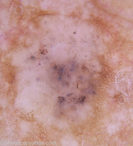
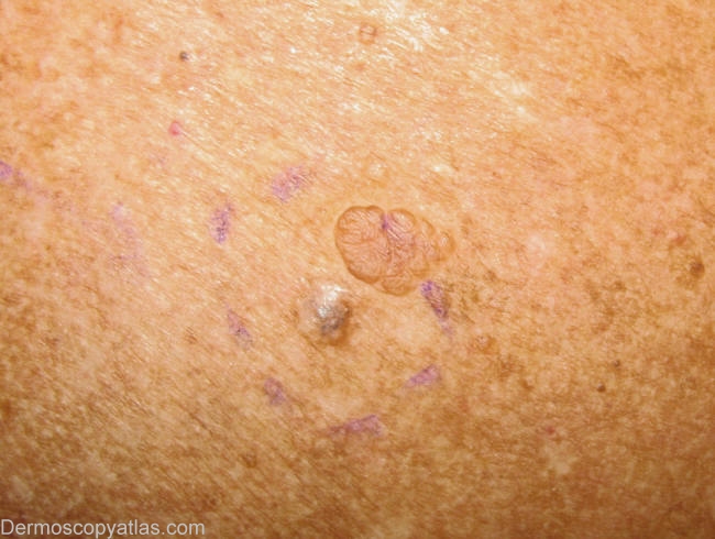
Description: Clinical- Papule.

History:

Presented for six monthly skin examination and melanoma follow-up.He has had Clark Level II melanoma excised from his mid upper back over two years ago.He also had few NMSCs excised from his lower back & face.This lesion was found during a routine examination.

Histopathology:  Basal cell carcinoma and benign,sessile intradermal naevus.

DermNetNZ   eMedicine   PubMed   Dermatology Online   Archives   JAAD for "BCC pigmented"

  • Recent Submissions
  • Search
  • Diagnosis
  • Quiz
  • Image comparison
  • Submit an Image
  • Teaching Cases
  • Index
  • About

Copyright 2022. Dermoscopy Atlas. ALL Rights Reserved.

Change Image