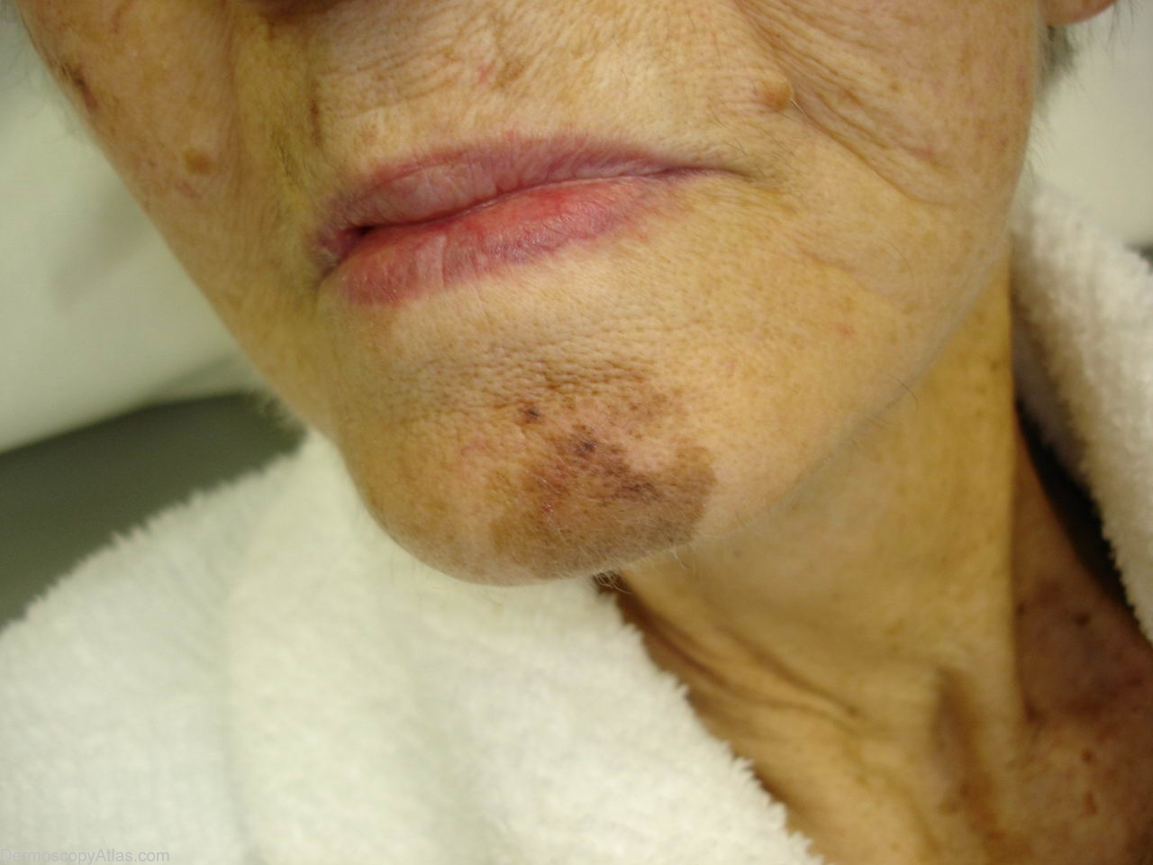

Site: Chin
Diagnosis: Lentigo Maligna
Sex: F
Age: 81
Type: Dermlite Polarised
Submitted By: Ian McColl
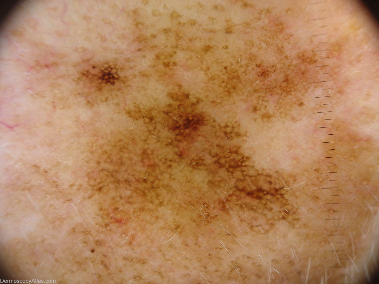
Description: Dermatoscopy of pigmented macule showing a thickened darker network in several areas.
History: She is in her 80s in reasonable general health and lives alone.I was asked to look at another problem she had but noted this lesion on her chin. I biopsied this in a couple of areas.
Clinically I thought this was a lentigo maligna. I biopsied the pigmented area at 10 o'clock and took a second punch from the uniformly brown area at 6 o'clock.
The pathology report for the upper area was as follows Sections show a biopsy of skin in which there is a proliferation of atypical melanocytes restricted to the basal epidermis. They form both a lentiginous as well as a nested pattern. There is no evidence of upward epidermal spread and no evidence of dermal involvement. The dermis shows marked solar elastosis. The features best fit for a dysplastic junctional lentiginous naevus. There is no evidence of malignancy.
The second biopsy was from the uniformly coloured area. It was reported as follows. Sections show a biopsy of skin in which there is a mild proliferation of atypical melanocytes in the basal epidermis. They form mainly a lentiginous pattern. No nests are present. The epidermis is atrophic and the underlying dermis shows marked solar elastosis. The degree of atypia in this biopsy is quite mild.
The pathologist reports the first biopsy as dysplastic lentiginous junctional nevus because of the nesting. He does not consider nesting a feature of lentigo maligna.He reports the second biopsy as lentigo maligna but the level of atypia is very mild and he might just as well have reported it as an atypical lentigine.He will not report a lesion as melanoma in situ unless there is some upward spread of atypical melanocytes into the epidermis. I have shown him the photographs of this lady's lesion. Clinically it is lentigo maligna. Dermoscopically it is lentigo maligna.The pathology is lagging behind at a morphological level. Soon some gene studies of these lesions may define them better but for the moment your eyes and the dermatoscope are better.
B
