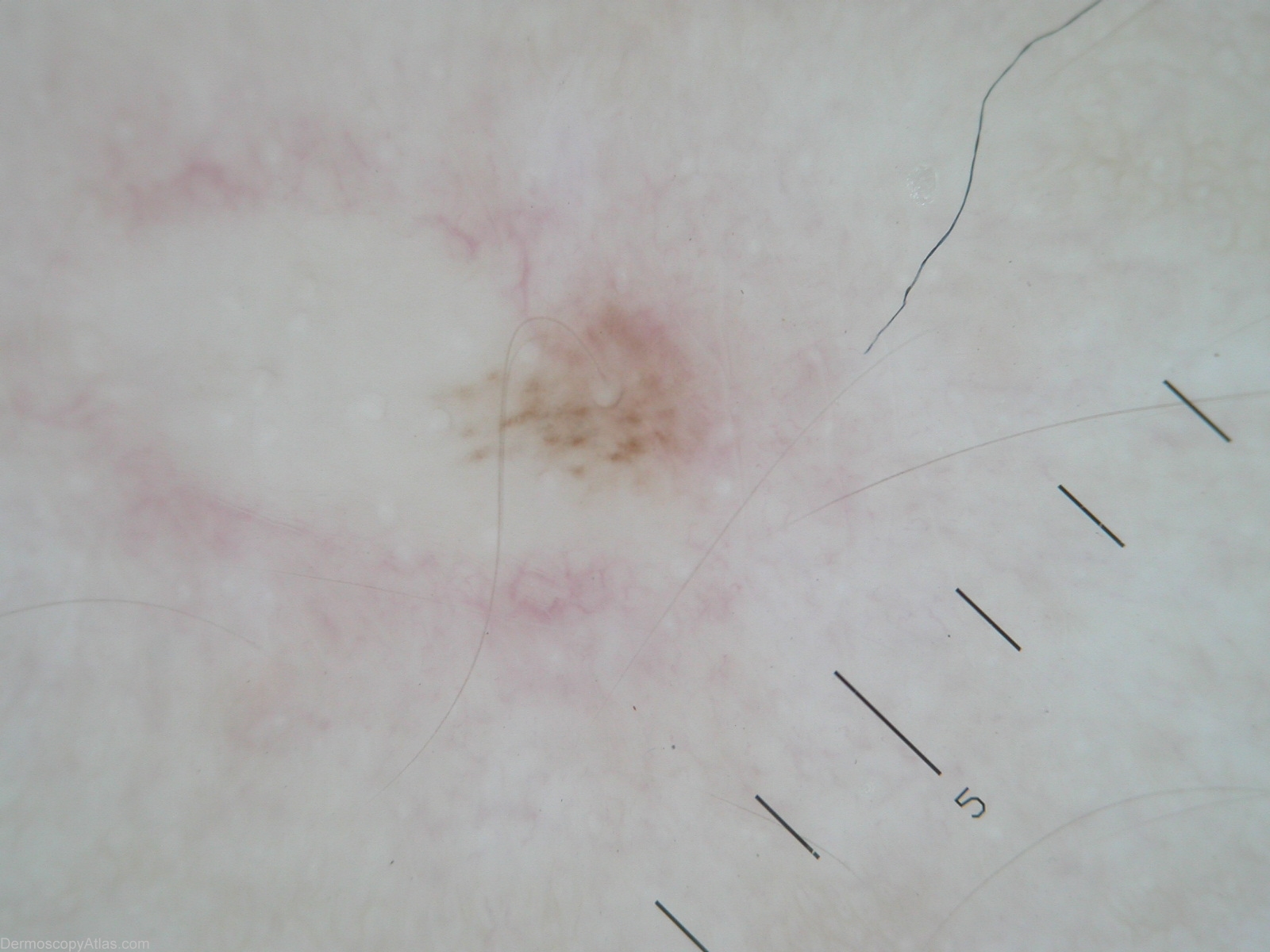
Loading
TeleDermConsult-logo
  • Recent Submissions
  • Search
  • Diagnosis
  • Quiz
  • Image comparison
  • Submit
  • Teaching Cases
  • Index
  • About

<< Back to Diagnosis Page

Image Number #2440 (Nevus recurrent)

Site: Back

Diagnosis: Nevus recurrent

Sex: F

Age: 26

Type: Heine

Submitted By: Stelios Minas

View Full Size Image

Description: Light brown pigmentation with some dots and globules

History:

The patient has a history of an excised pigmented lesion done with laser a 2 years ago. Some time after this she has developed a new lesion precisely at the excision site. No previous histopathology report (laser excision).

Laser excision is not recommended  for melanocytic lesions.

Dermoscopic description: Light brown pigmentation with some dots and globules. 

As Ian McColl said:Always check the original histopathology of a previous excision.  In benign recurrences the pigment usually does not extend beyond the scar margins.

DermNetNZ   eMedicine   PubMed   Dermatology Online   Archives   JAAD for "Nevus recurrent"

  • Recent Submissions
  • Search
  • Diagnosis
  • Quiz
  • Image comparison
  • Submit an Image
  • Teaching Cases
  • Index
  • About

Copyright 2022. Dermoscopy Atlas. ALL Rights Reserved.

Change Image