Loading
TeleDermConsult-logo
  • Recent Submissions
  • Search
  • Diagnosis
  • Quiz
  • Image comparison
  • Submit
  • Teaching Cases
  • Index
  • About

Teaching Cases


teach1

History

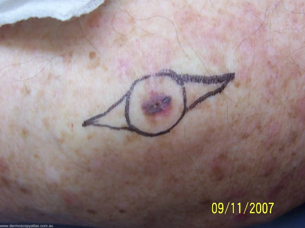
Case 9 Male age 72. Routine check. Famoly concerned about a lesion on the R calf.

teach1

View Full Size Image

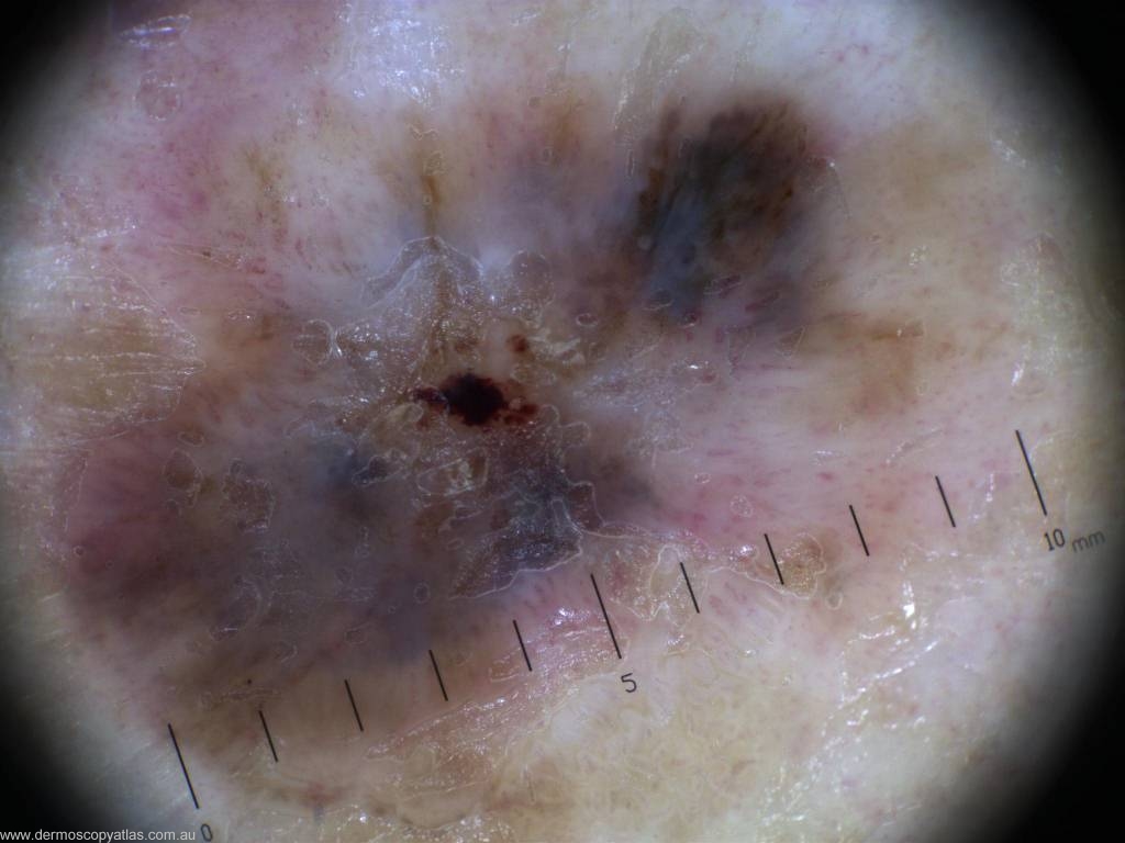
Question: What is the diagnosis? Consider Seborrhoeic keratosis, Junctional nevus, Invasive melanoma, Pigmented BCC, Dysplastic nevus and Pigmented IEC.

Answer: Histology: L2 0.7mm thick, superficial spreading melanoma.


Next Question
  • Recent Submissions
  • Search
  • Diagnosis
  • Quiz
  • Image comparison
  • Submit an Image
  • Teaching Cases
  • Index
  • About

Copyright 2022. Dermoscopy Atlas. ALL Rights Reserved.

Change Image