
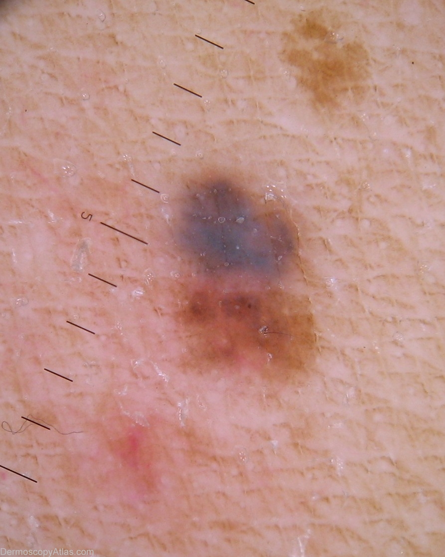
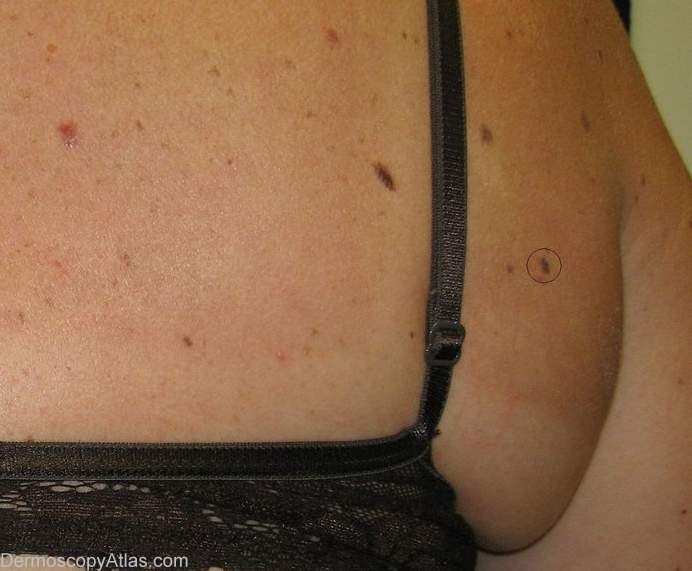
Site: Axillae
Diagnosis: Nevus combined
Sex: F
Age: 46
Type: Heine
Submitted By: Ian McColl
Description: Heine view of lesion in axillae. There are two cell populations here shown by the blue and brown areas. The blue area is suggestive of a blue grey veil.
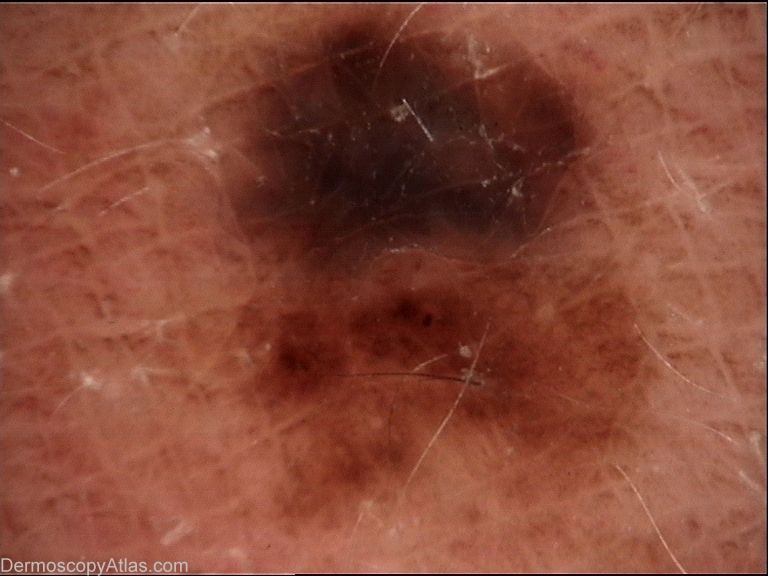
History: This is a combined nevus on histology. The Molemax image does not show the blue of the Heine.The histology report was as follows Right Posterior Axillary Fold: The sections show a predominantly
intradermal, compound COMBINED NAEVUS, which comprises conventional
acquired type compound naevus and a Paget cell type epithelioid naeval
component. The paget naevomelanocytes are characterised by large cell
size, voluminous pale cytoplasm finely dusted with melanin and mild
nuclear variability. A mild chronic inflammatory cell infiltrate mainly
comprising melanophages accompanies the latter component. No dysplastic
naevus features are identified. Punch margins appear clear of naevus in
the plane of section examined.
No evidence of malignancy/melanoma is seen.
PUNCH EXCISION, RIGHT POSTERIOR AXILLARY FOLD
COMPOUND COMBINED NAEVUS. PUNCH MARGINS APPEAR CLEAR.
View a Blog discussion of this case
View a recent JAAD letter on the histology of the Combined nevus