Loading
TeleDermConsult-logo
  • Recent Submissions
  • Search
  • Diagnosis
  • Quiz
  • Image comparison
  • Submit
  • Teaching Cases
  • Index
  • About

<< Back to Diagnosis Page

Image Number #3849 (Melanoma nodular)

Site: Abdomen

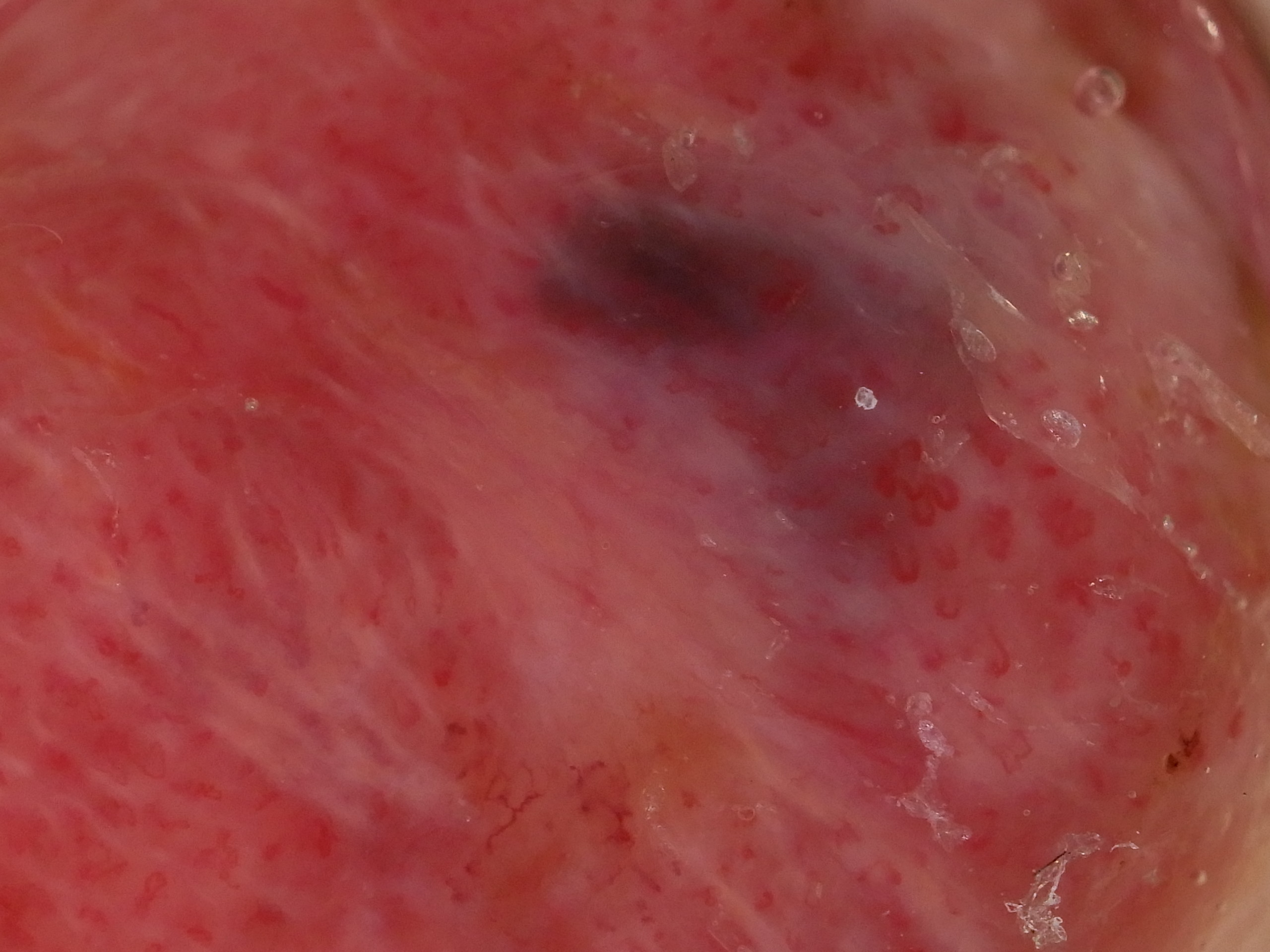
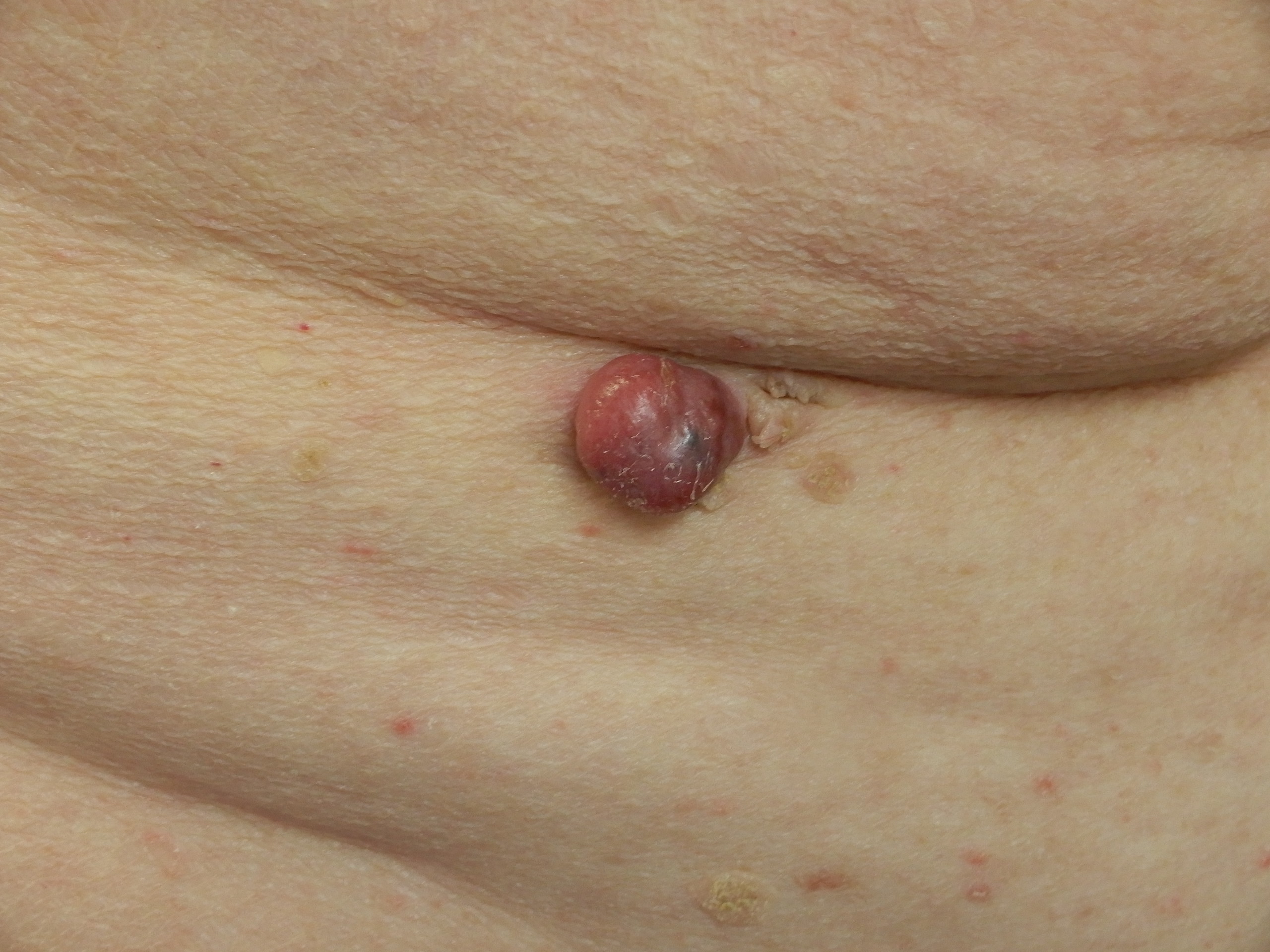
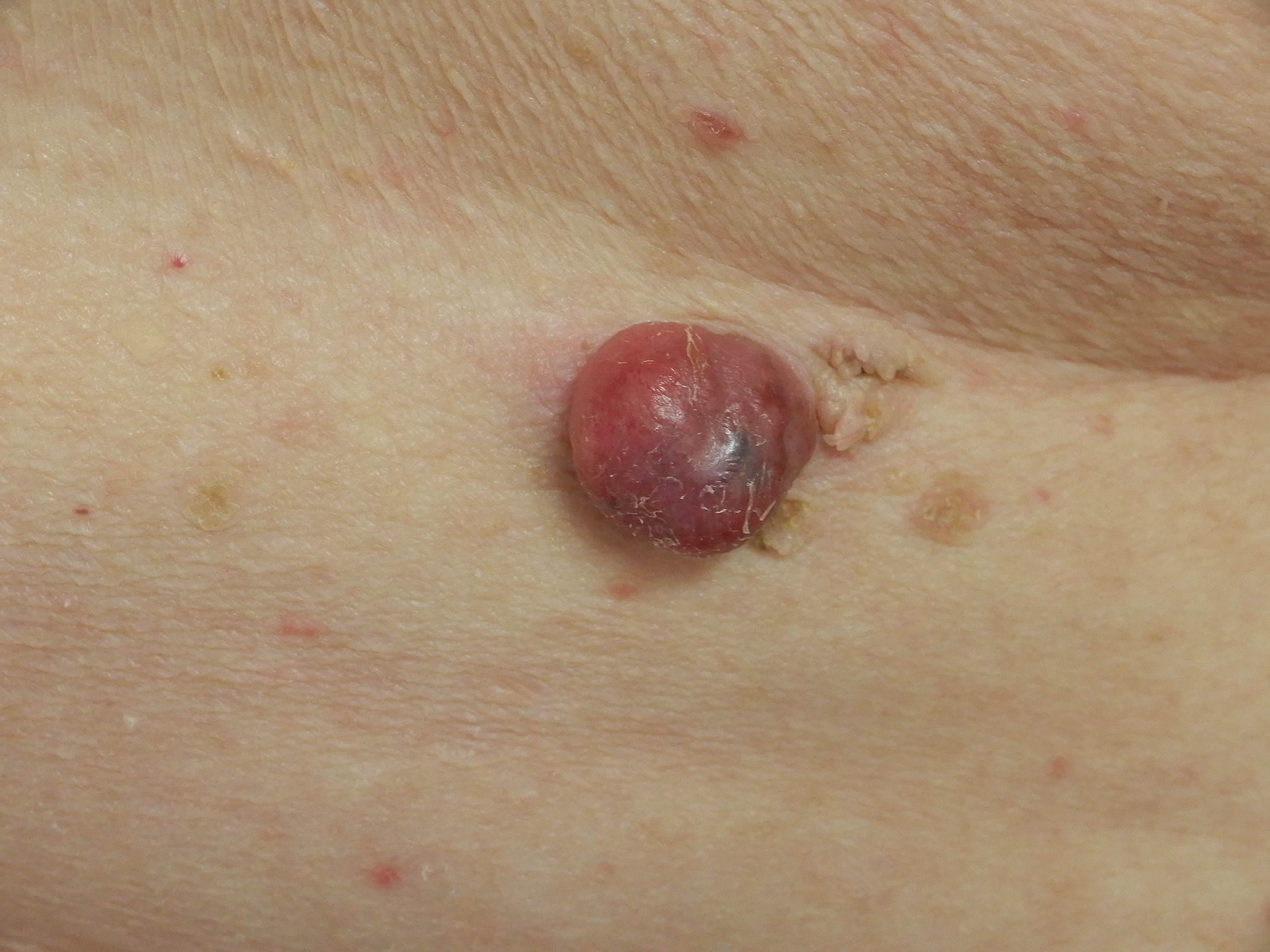
Diagnosis: Melanoma nodular

Sex: M

Age: 74

Type: Dermlite Polarised

Submitted By: Ian McColl

View Full Size Image

Click Here to View Details

Description:

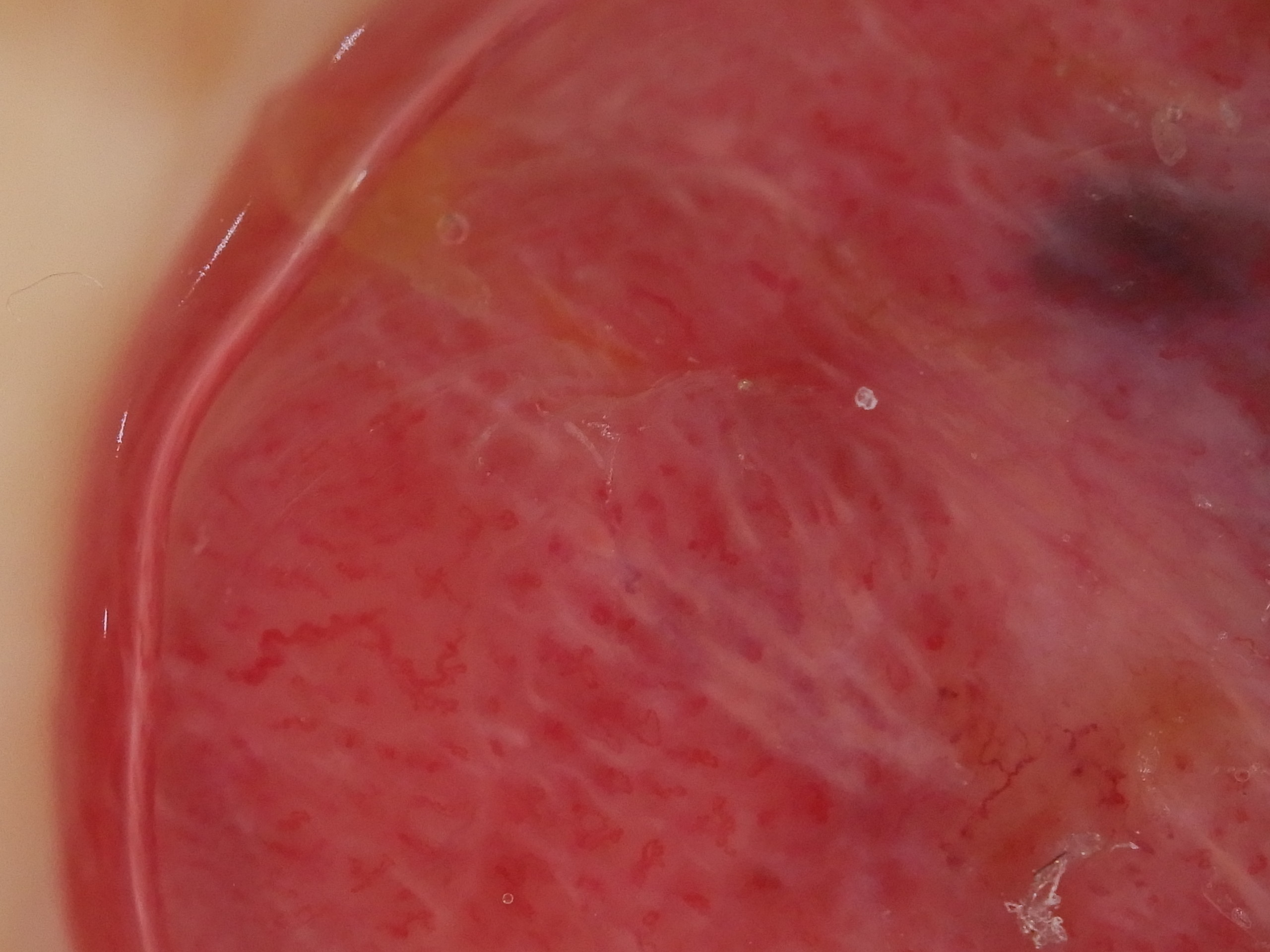
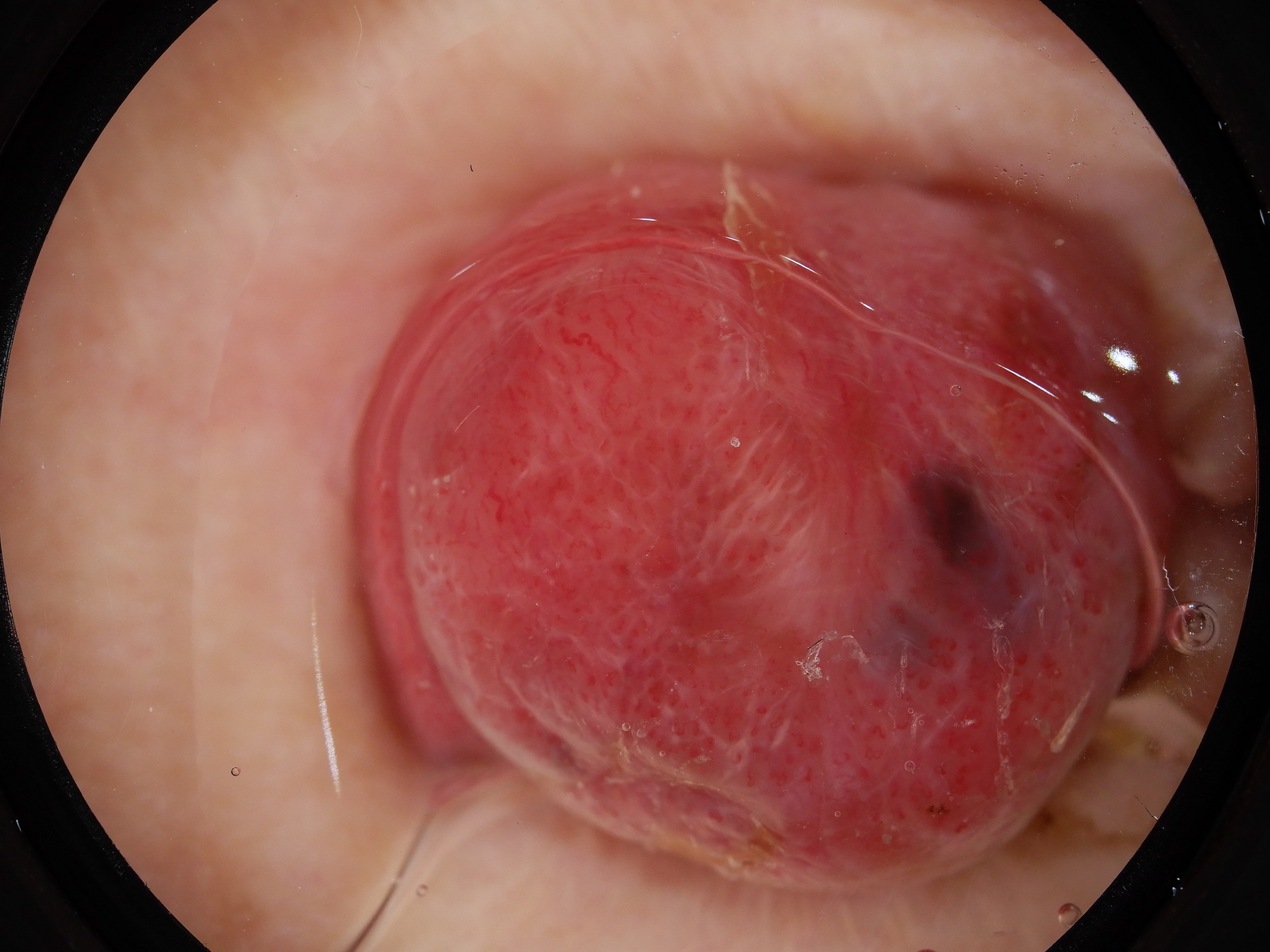
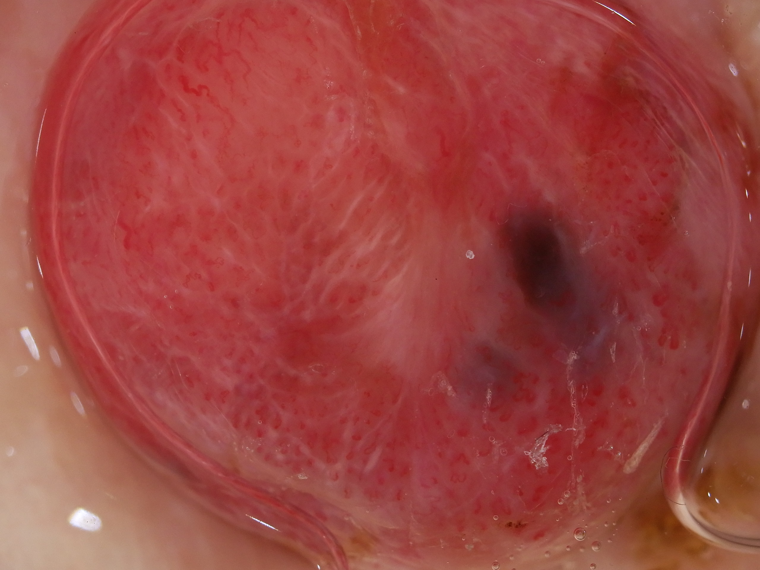
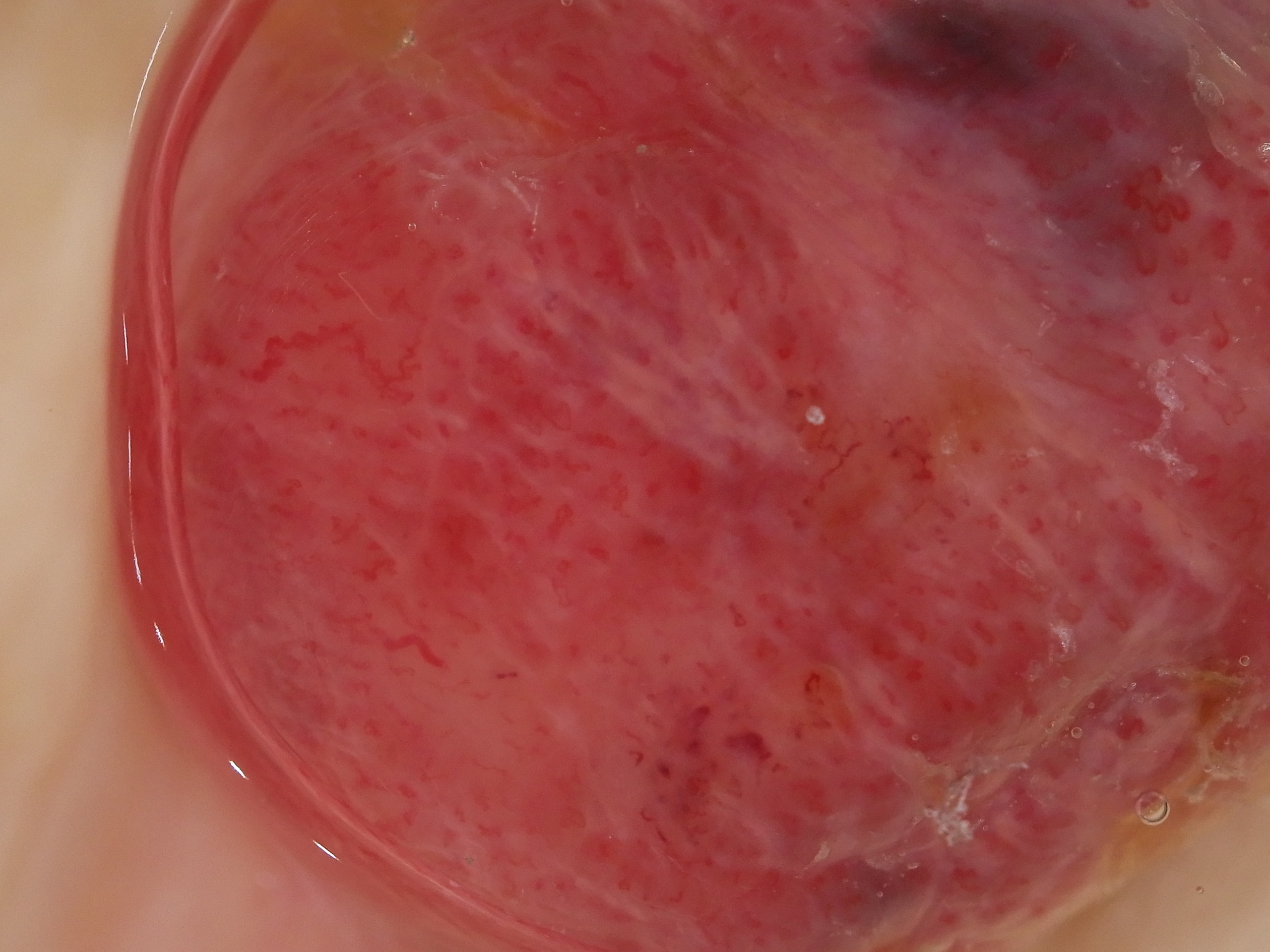
Pink nodule with white lines and pigmented clods

History:

This nodule grew rapidly over a 6 week period with no history of a pre existing lesion. The patient had no past history of melanoma. The lesion was painless.

It was a 7 mm thick nodular melanoma with many mitoses. 

DermNetNZ   eMedicine   PubMed   Dermatology Online   Archives   JAAD for "Melanoma nodular"

  • Recent Submissions
  • Search
  • Diagnosis
  • Quiz
  • Image comparison
  • Submit an Image
  • Teaching Cases
  • Index
  • About

Copyright 2022. Dermoscopy Atlas. ALL Rights Reserved.

Change Image