
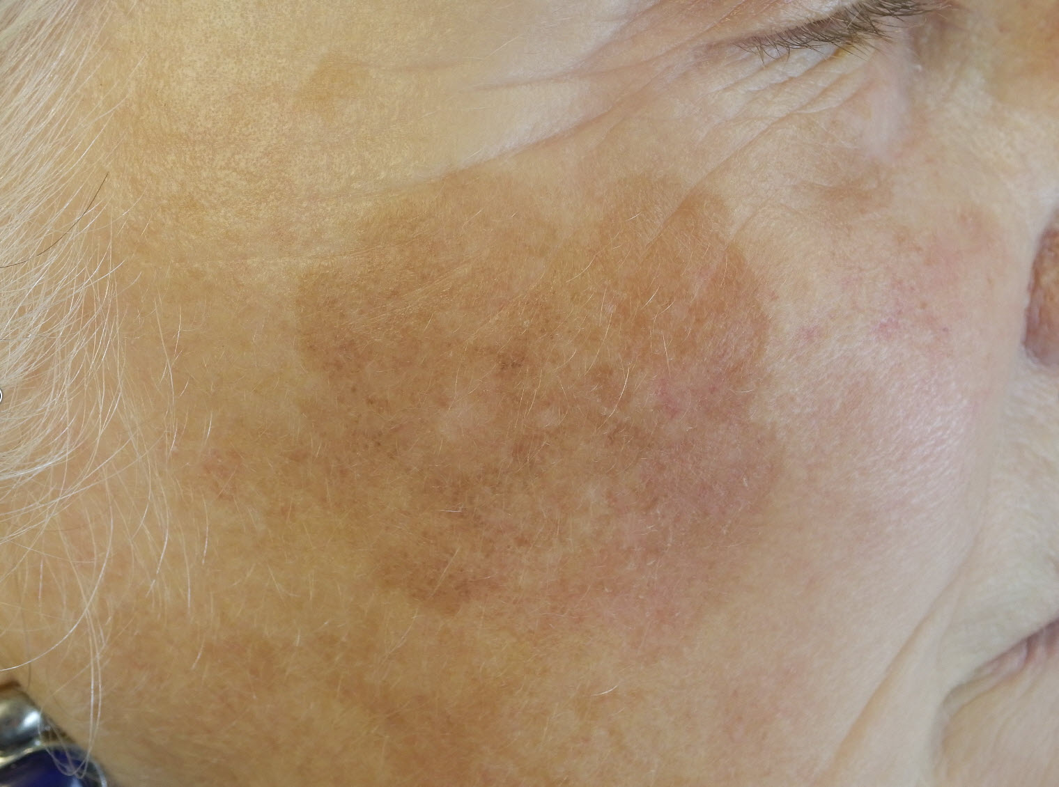
Site: Cheek
Diagnosis: Large cell acanthoma
Sex: F
Age: 71
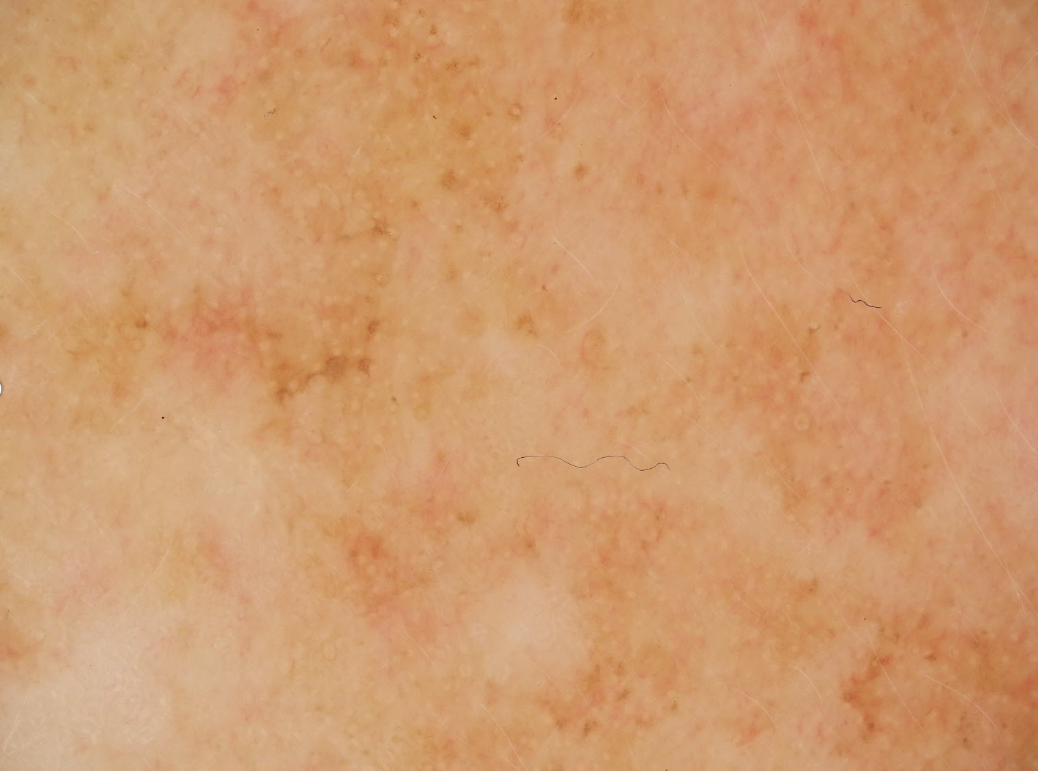
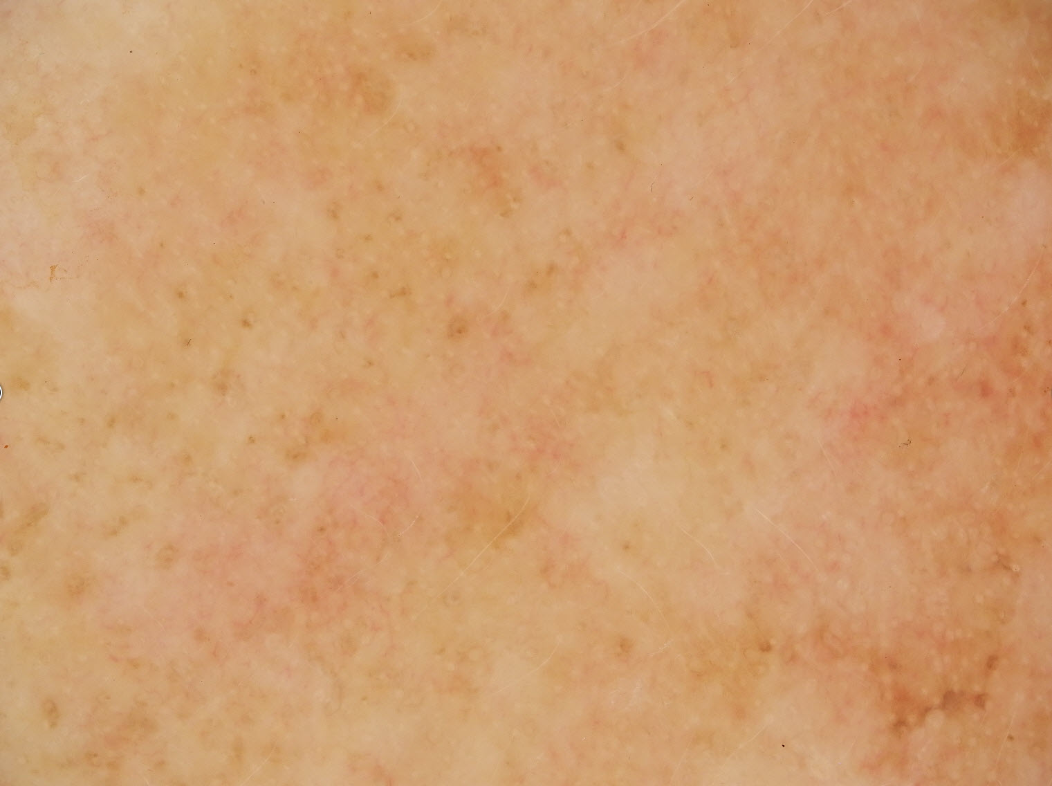
Type: Dermlite Polarised
Submitted By: Ian McColl
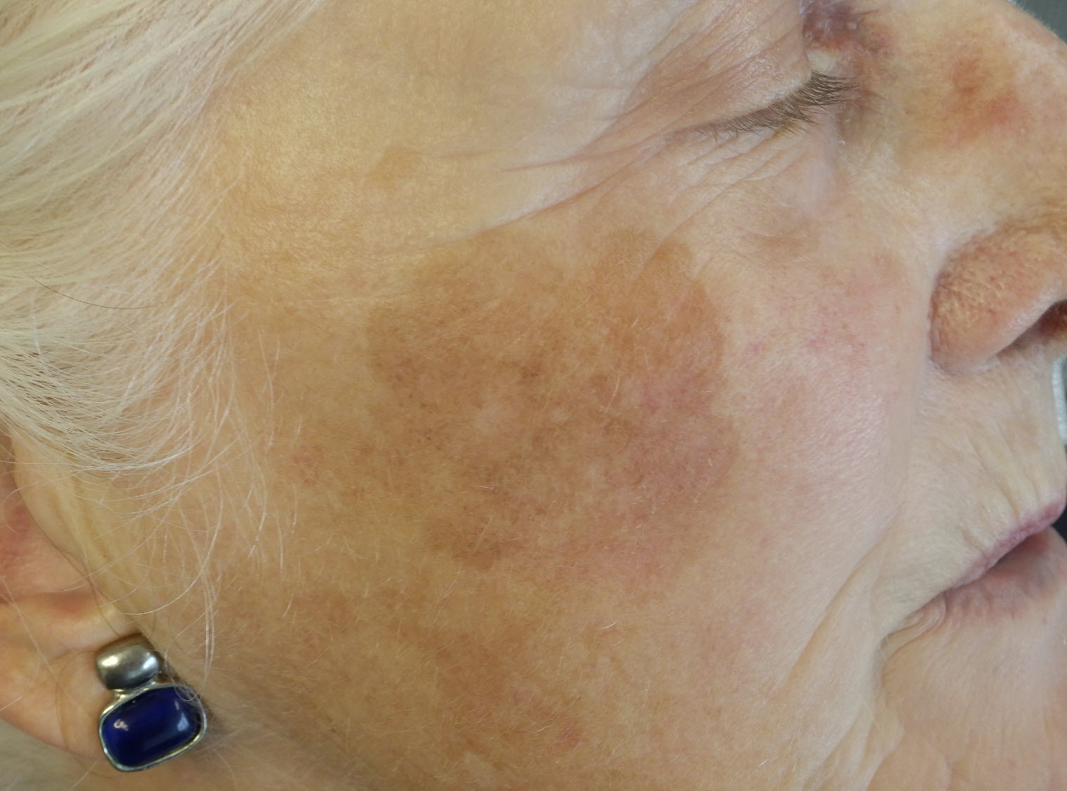
Description: Large pigmented lesion cheek
History:
Case 1 This lesion has been slowly growing over 15 years. What do you think? Worth biopsying or just keep letting it grow?
I biopsied in 4 areas but no atypical melanocytic proliferation. It was reported as a large cell acanthoma ie a type of seb k. I could treat it with a scanning CO2 laser if she wants.
Dermatoscopically the issue was whether she had any grey circles that would have suggested lentigo maligna. Several shaves here might have been a better option for the biopsy.