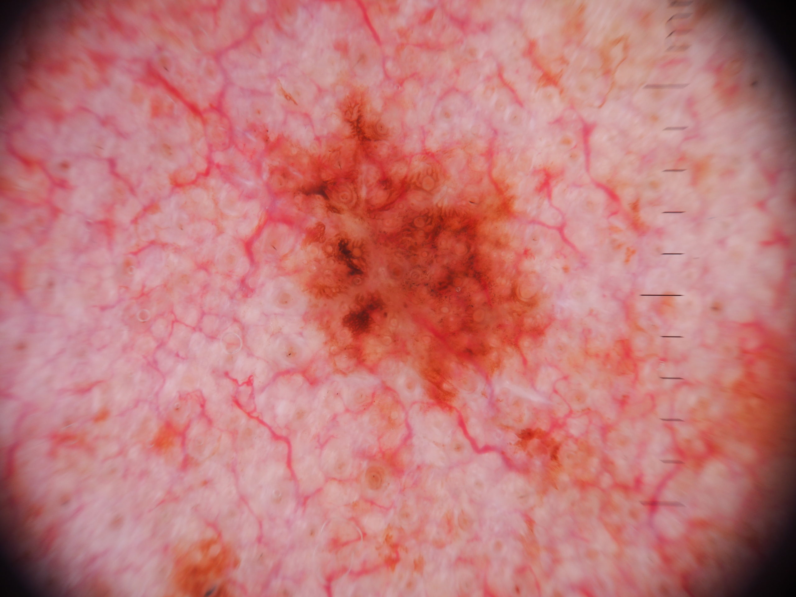
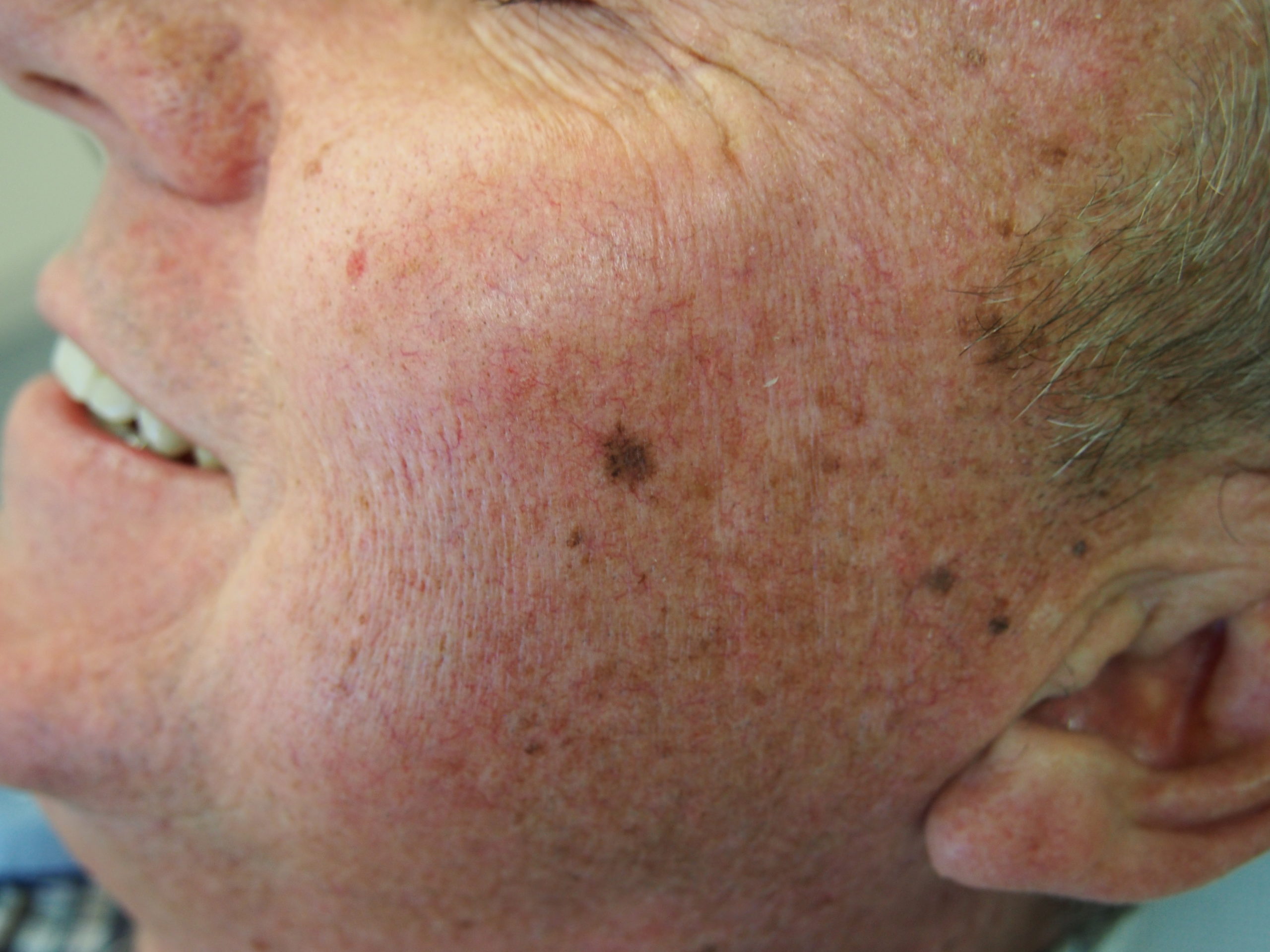
Loading
TeleDermConsult-logo
  • Recent Submissions
  • Search
  • Diagnosis
  • Quiz
  • Image comparison
  • Submit
  • Teaching Cases
  • Index
  • About

<< Back to Diagnosis Page

Image Number #3804 (Lentigo Maligna)

Site: Cheek

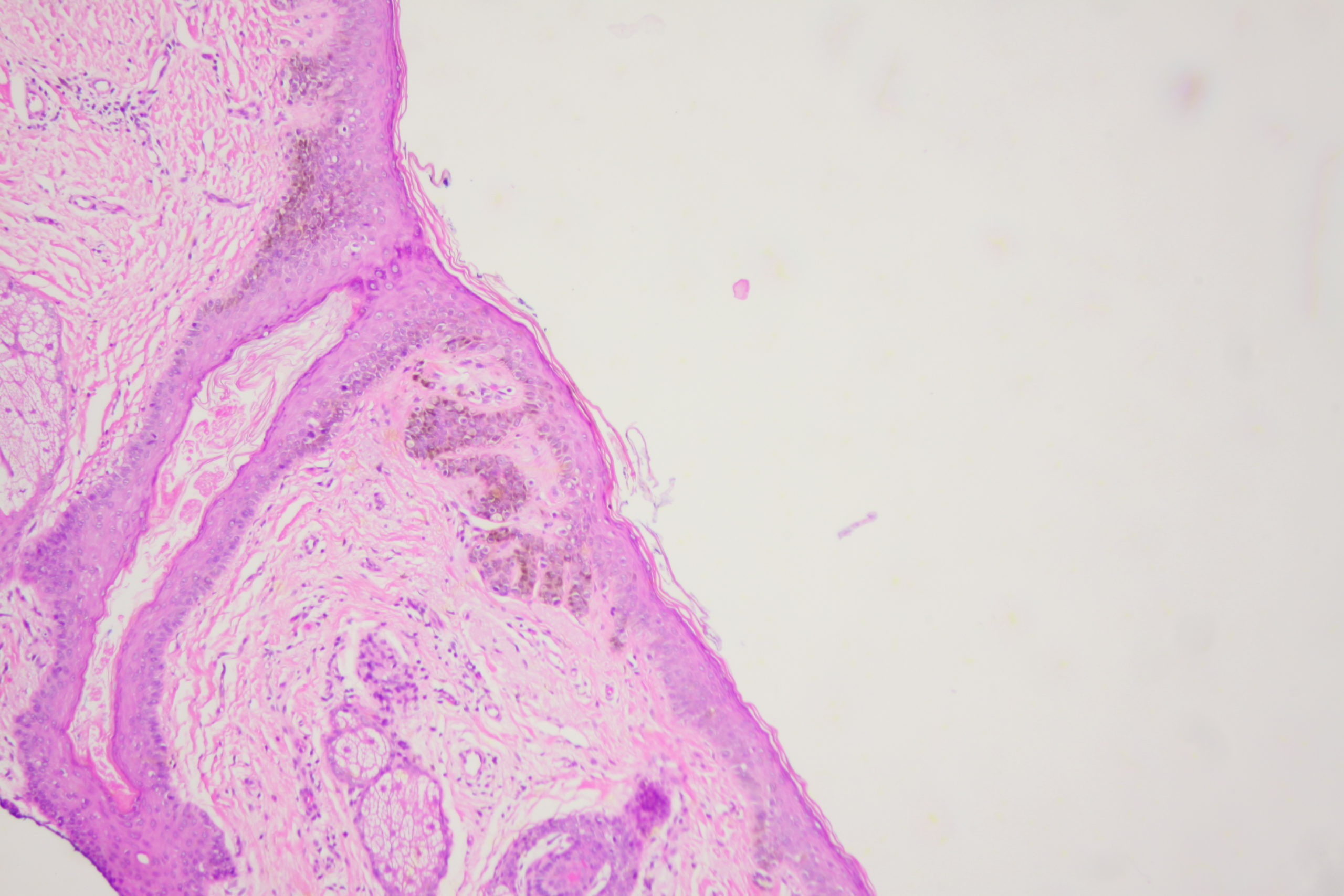
Diagnosis: Lentigo Maligna

Sex: M

Age: 65

Type: Dermlite Polarised

Submitted By: Ian McColl

View Full Size Image

Click Here to View Details

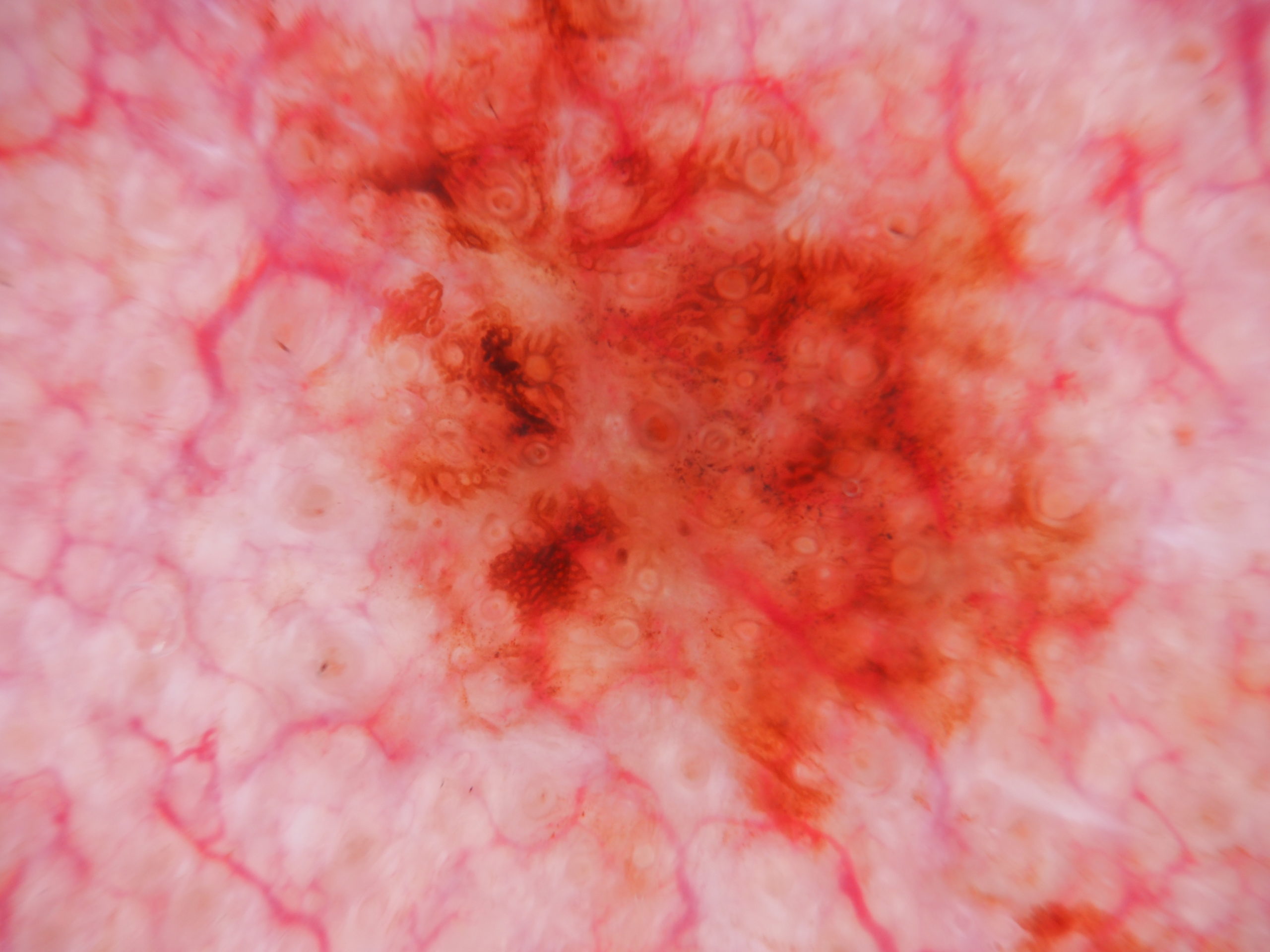
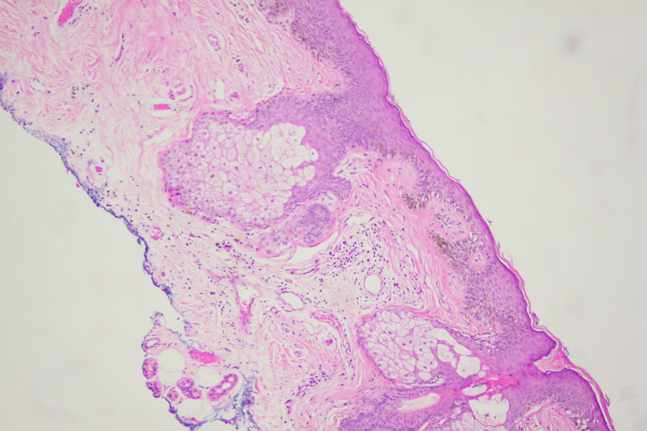
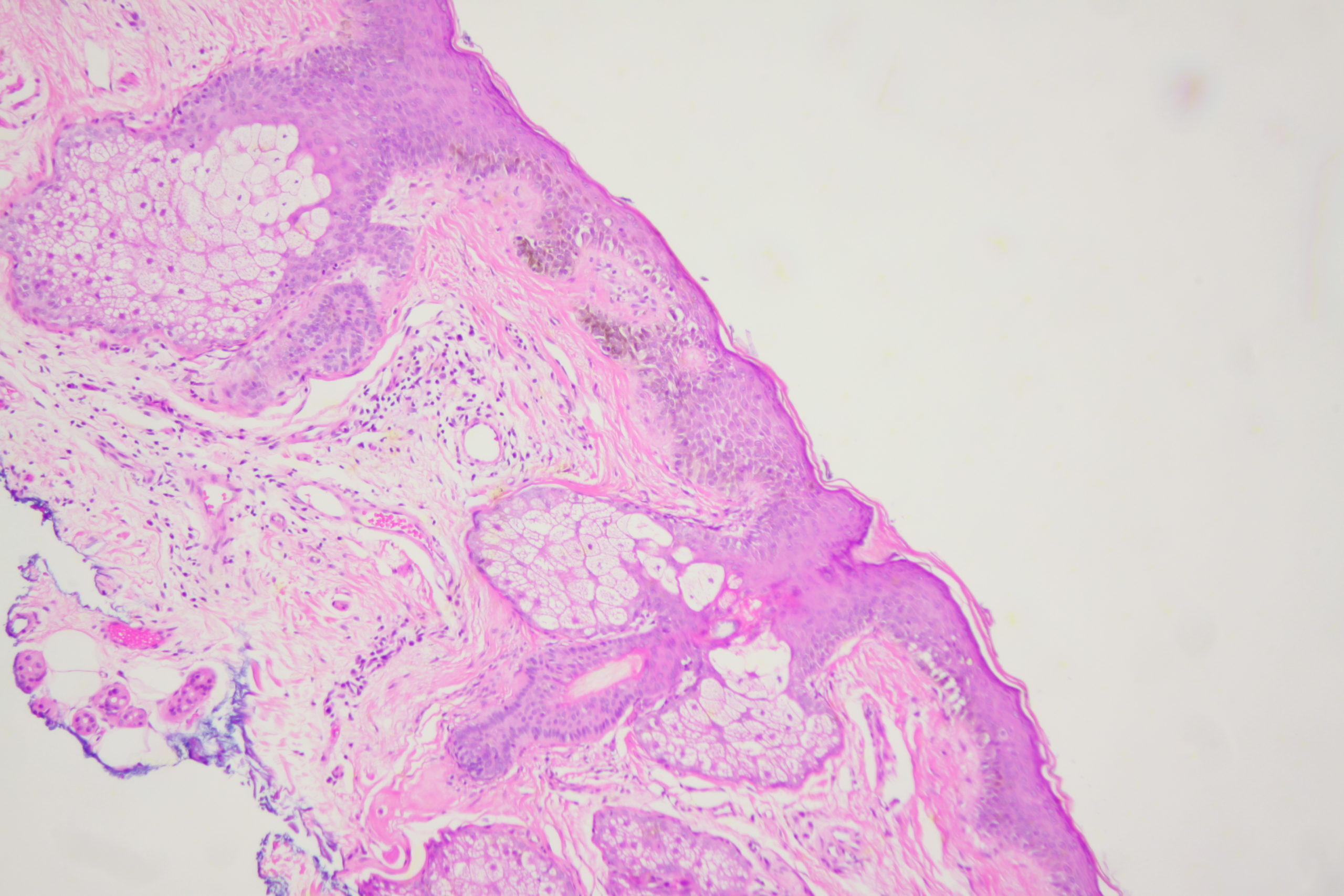
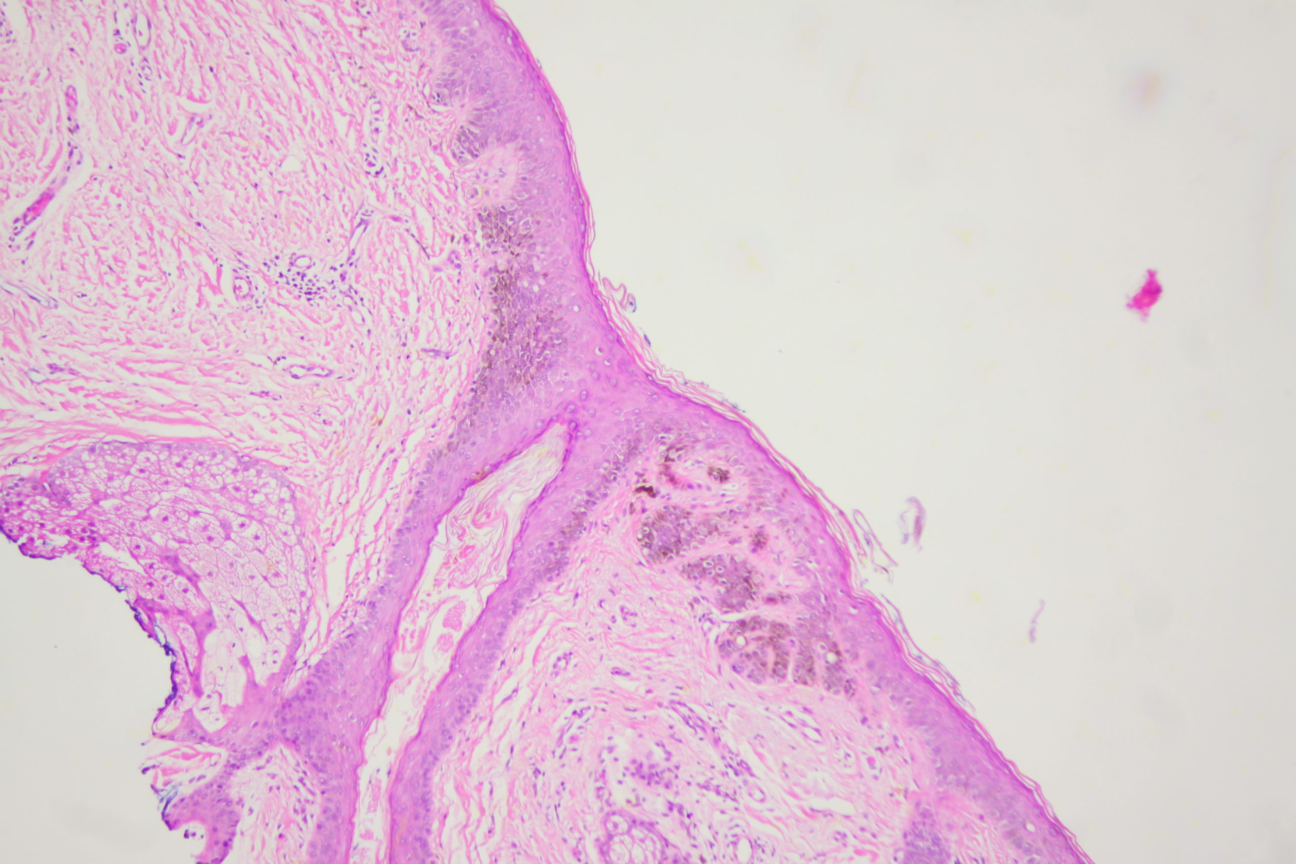
Description: Pigment dots and grey circles

History:

A pigmented lesion on the cheek that has slowly been increasing in size over several years. Male aged 65. Smooth surface.

Dermatoscopy shows pigment dots and pigment around follicles as grey circles. PRAME and Sox 10 showed atypical melanocytes along the DEJ extending into and down follicles with localised upward pagetoid spread. Early Lentigo maligna / Superficial intraepidermal melanoma.

DermNetNZ   eMedicine   PubMed   Dermatology Online   Archives   JAAD for "Lentigo Maligna"

  • Recent Submissions
  • Search
  • Diagnosis
  • Quiz
  • Image comparison
  • Submit an Image
  • Teaching Cases
  • Index
  • About

Copyright 2022. Dermoscopy Atlas. ALL Rights Reserved.

Change Image