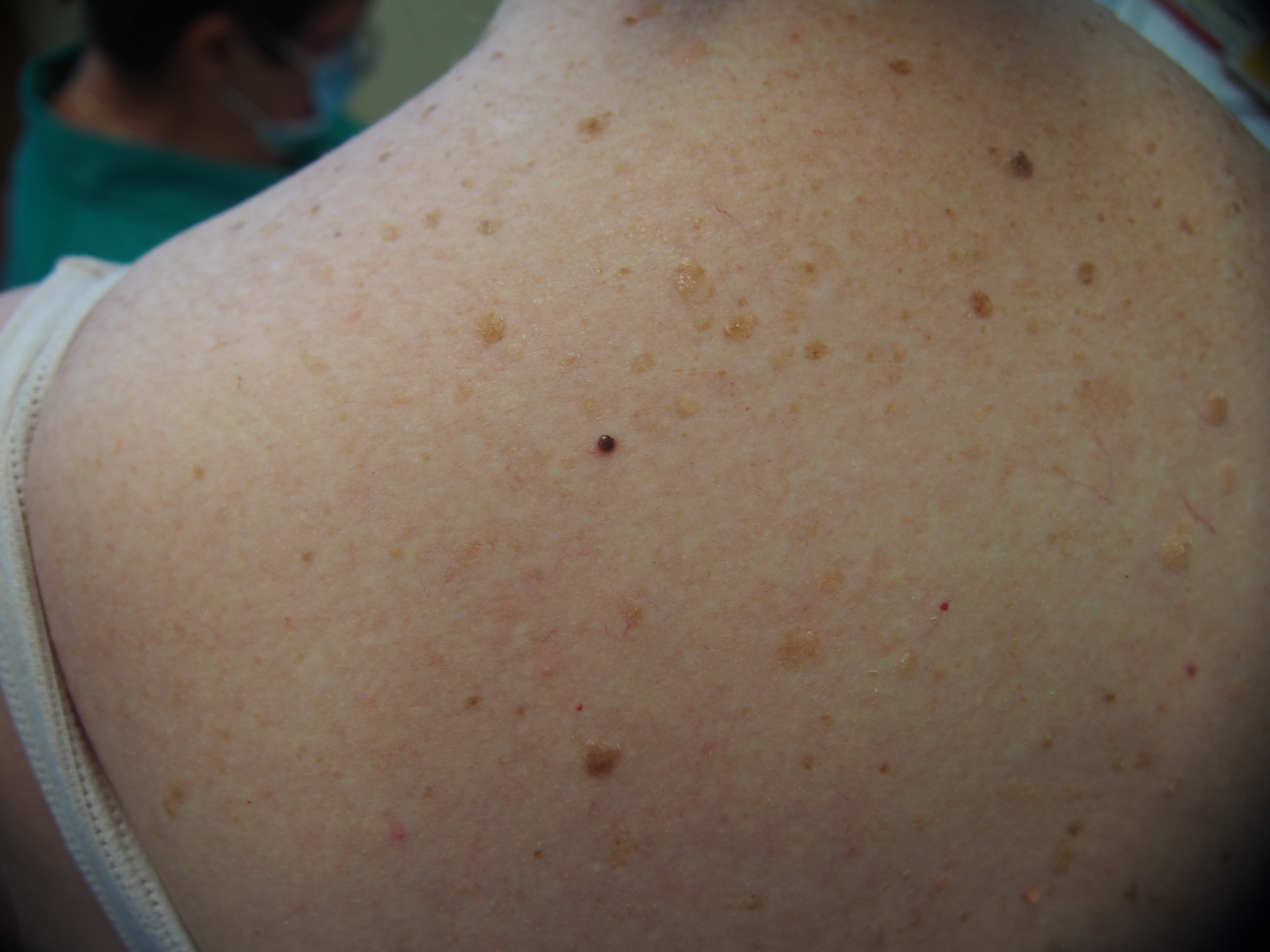
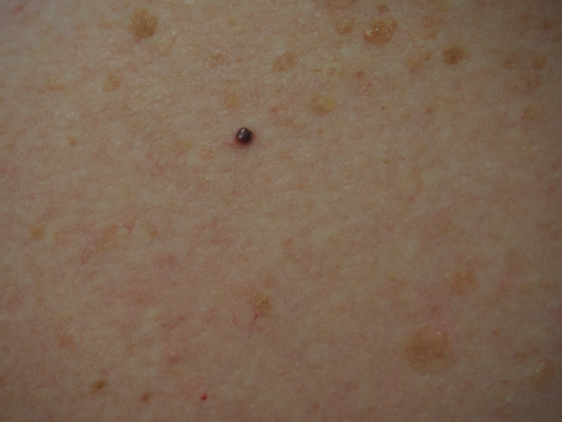
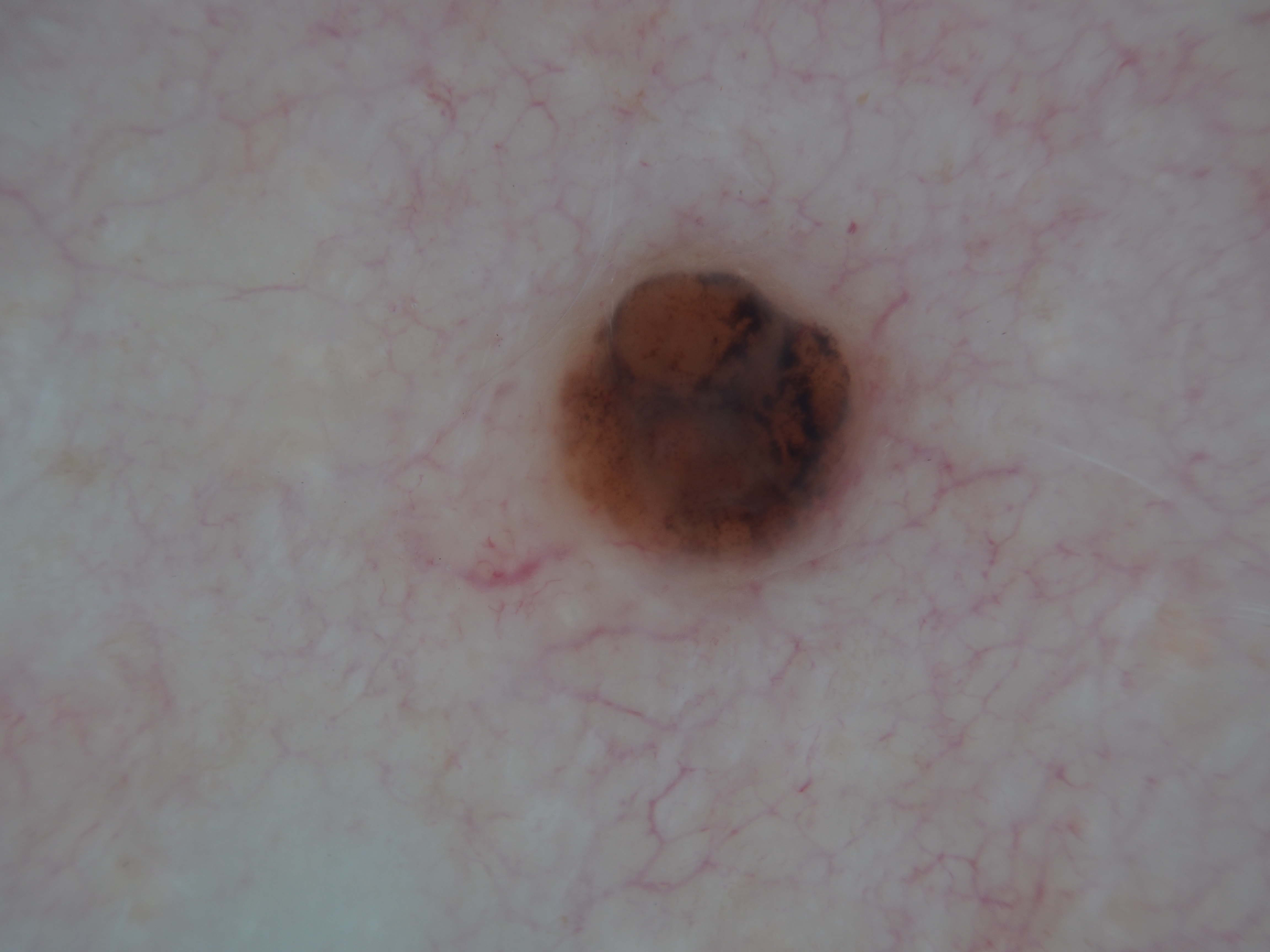
Loading
TeleDermConsult-logo
  • Recent Submissions
  • Search
  • Diagnosis
  • Quiz
  • Image comparison
  • Submit
  • Teaching Cases
  • Index
  • About

<< Back to Diagnosis Page

Image Number #3769 (Melanoma nodular)

Site: Back

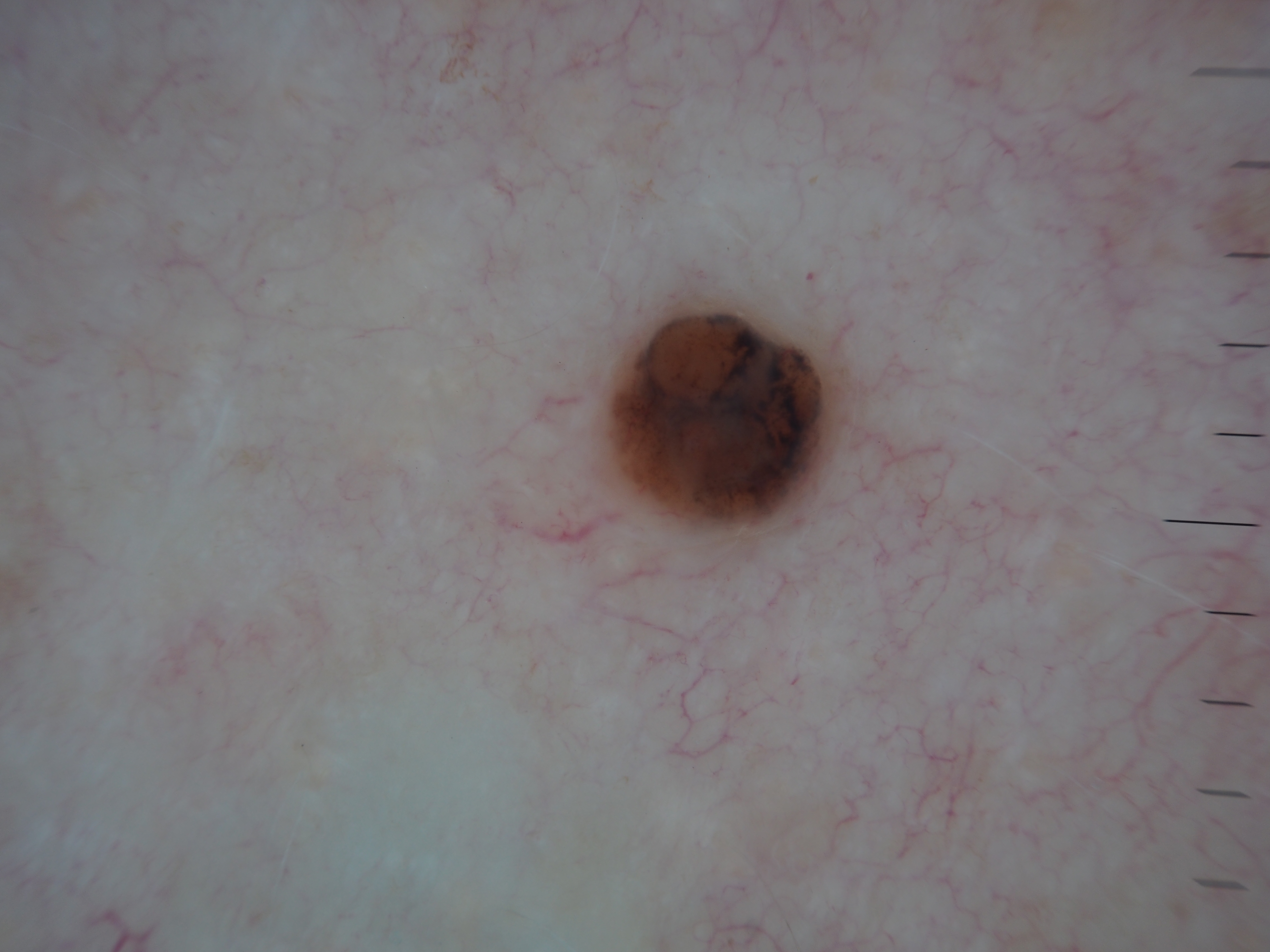
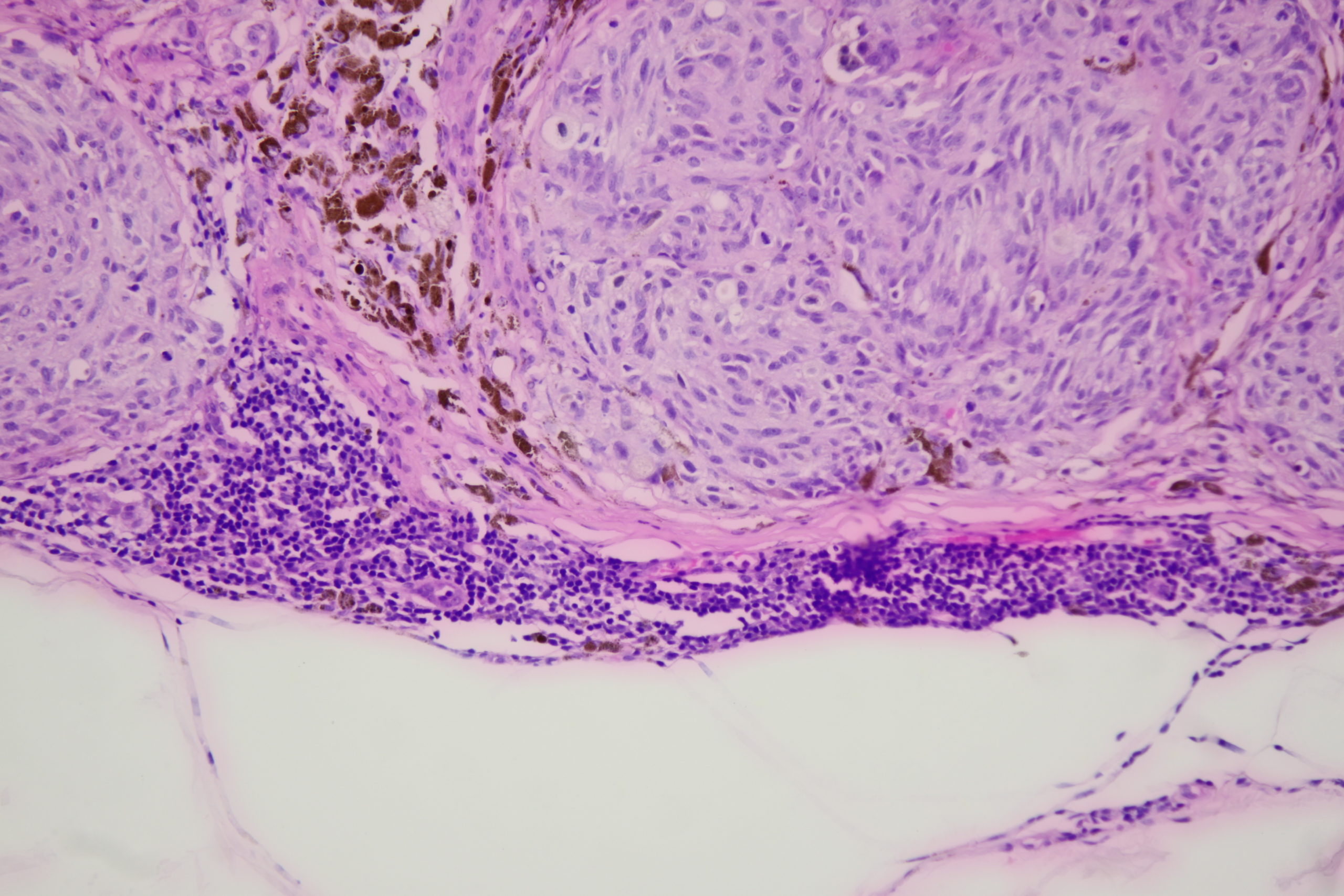
Diagnosis: Melanoma nodular

Sex: F

Age: 67

Type: Dermlite Polarised

Submitted By: Ian McColl

View Full Size Image

Click Here to View Details

Description:

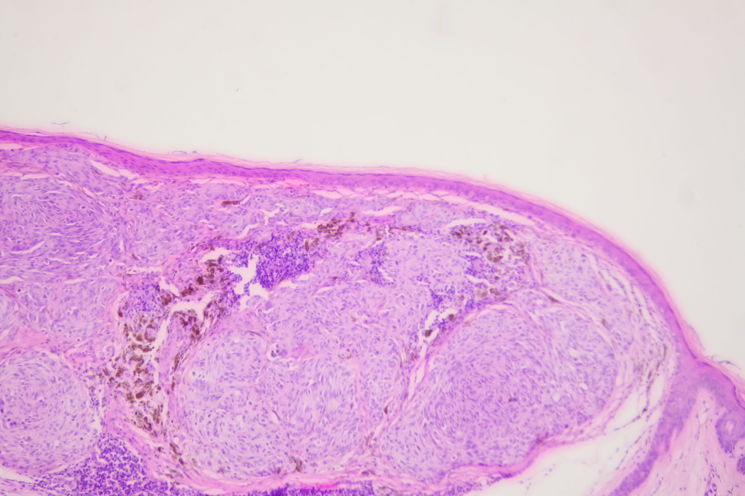
Spindled melanocytes in the dermis, No epidermal involvement

History:

This lesion was noted during a routine skin examination in an elderly patient on immunosuppressives for an autoimmune disease.

The lesion was a 0.55 mm thick Clark level 3 nodular melanoma.

See Nodular Melanoma in Dermoscopy Made Simple

DermNetNZ   eMedicine   PubMed   Dermatology Online   Archives   JAAD for "Melanoma nodular"

  • Recent Submissions
  • Search
  • Diagnosis
  • Quiz
  • Image comparison
  • Submit an Image
  • Teaching Cases
  • Index
  • About

Copyright 2022. Dermoscopy Atlas. ALL Rights Reserved.

Change Image