

Site: Shoulder
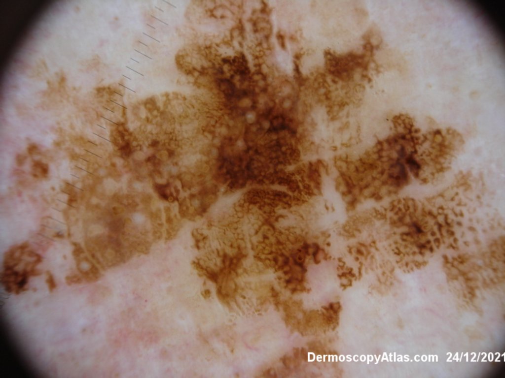
Diagnosis: Kittler Seborrhoeic keratosis reticulate
Sex: M
Age: 63
Type: Dermlite Polarised
Submitted By: Ian McColl

Description: Clinical
History:
Seborrhoeic keratoses are great dermatoscopic imitators. Usually you see crypts or clods of various colours depending on where the keratin is. The clods can be orange, yellow, black, brown or white. The background pigmentation of a seb k can be minimal (pink) lesion or jet black as in the so called melanoacanthoma.
View Seb K in Dermoscopy Made Simple
Flat thin lesions can look like a solar lentigo with lines retic or curved and with brown circles on the face with pigment around the openings of hair follicles.
Lines curved of varying thickness sometimes showing a parallel orientation are seen in thicker lesions but they also tend to show various coloured clods