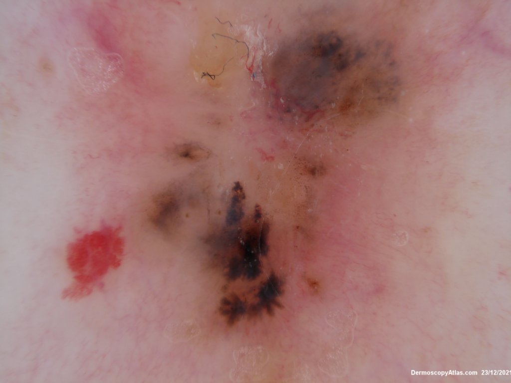
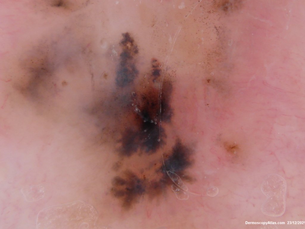
Loading
TeleDermConsult-logo
  • Recent Submissions
  • Search
  • Diagnosis
  • Quiz
  • Image comparison
  • Submit
  • Teaching Cases
  • Index
  • About

<< Back to Diagnosis Page

Image Number #3614 (Kittler Basal cell carcinoma)

Site: Back

Diagnosis: Kittler Basal cell carcinoma

Sex: M

Age: 63

Type: Heine

Submitted By: Ian McColl

View Full Size Image

Click Here to View Details

Description:

History:

 A pigmented BCC can show as here grey dots, lines peripheral radial meeting at a point, blue grey structures, ulceration, some brown dots and linear focussed branching vessels. Blue clods are also a feature of pigmented BCC but they are not well shown here.

See dermatoscopic features of pigmented BCC in Dermoscopy Made Simple

DermNetNZ   eMedicine   PubMed   Dermatology Online   Archives   JAAD for "Kittler Basal cell carcinoma"

  • Recent Submissions
  • Search
  • Diagnosis
  • Quiz
  • Image comparison
  • Submit an Image
  • Teaching Cases
  • Index
  • About

Copyright 2022. Dermoscopy Atlas. ALL Rights Reserved.

Change Image