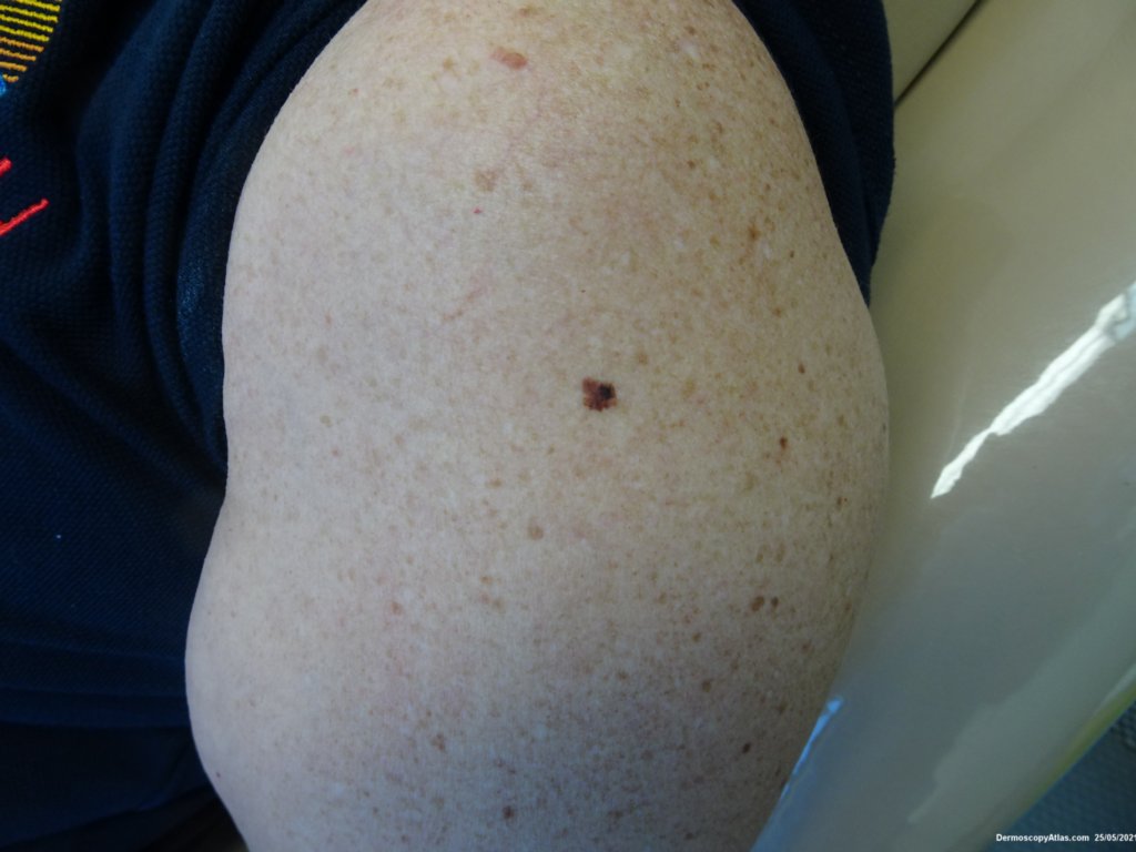
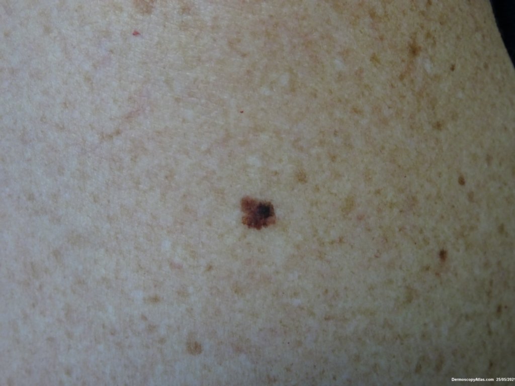
Loading
TeleDermConsult-logo
  • Recent Submissions
  • Search
  • Diagnosis
  • Quiz
  • Image comparison
  • Submit
  • Teaching Cases
  • Index
  • About

<< Back to Diagnosis Page

Image Number #3591 (Melanoma invasive)

Site: Arm,upper

Diagnosis: Melanoma invasive

Sex: F

Age: 42

Type: Dermlite Polarised

Submitted By: Ian McColl

View Full Size Image

Click Here to View Details

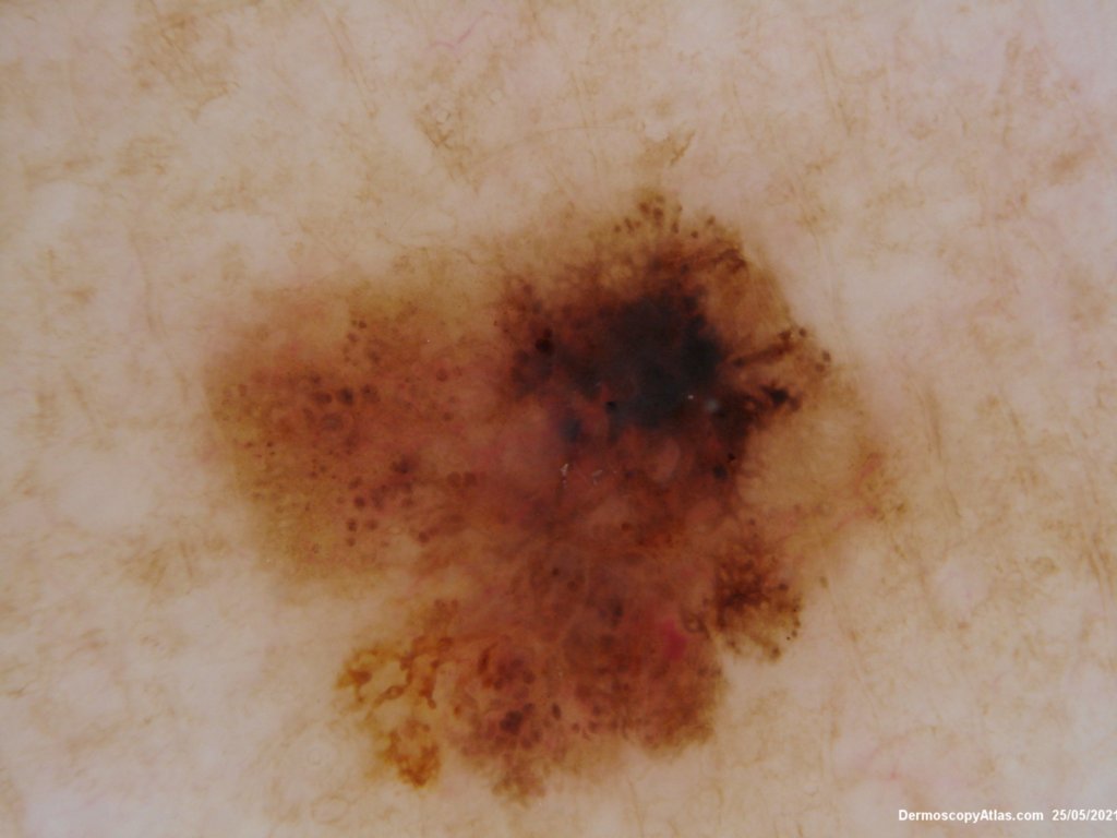
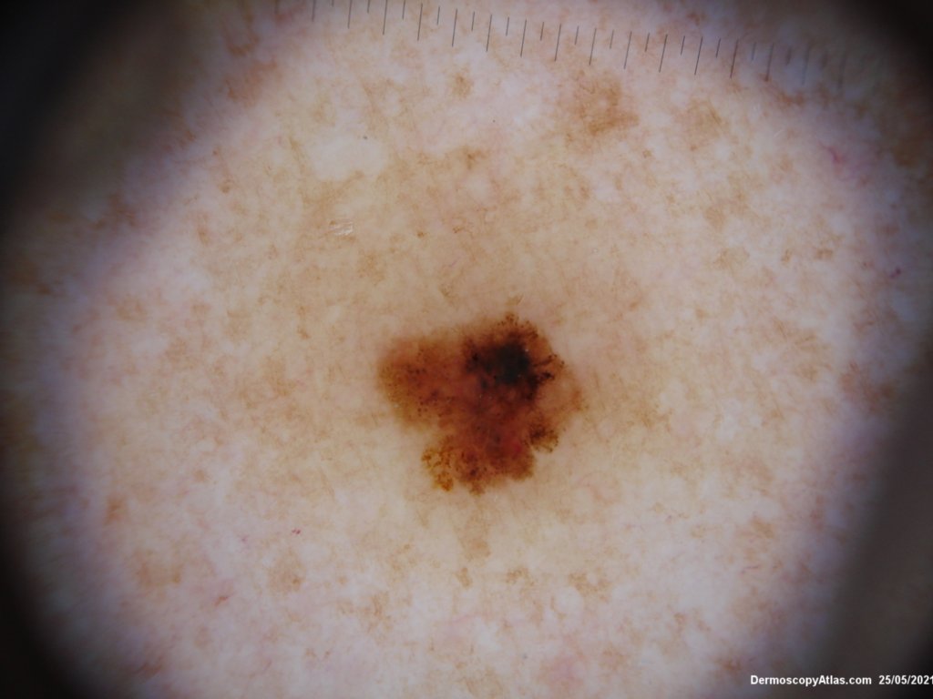
Description: Pigmented lesion with lines radial peripheral

History:

 Lady in her 40s with two previous in situ melanomas. This one was invasive 0.4 mm but her prognosis is still excellent. 

Dermatoscopically there were lines radial peripheral parallel, a few pseudopods, dark dots and thickened lines reticular. 

For review see Dermoscopy Made Simple 

DermNetNZ   eMedicine   PubMed   Dermatology Online   Archives   JAAD for "Melanoma invasive"

  • Recent Submissions
  • Search
  • Diagnosis
  • Quiz
  • Image comparison
  • Submit an Image
  • Teaching Cases
  • Index
  • About

Copyright 2022. Dermoscopy Atlas. ALL Rights Reserved.

Change Image