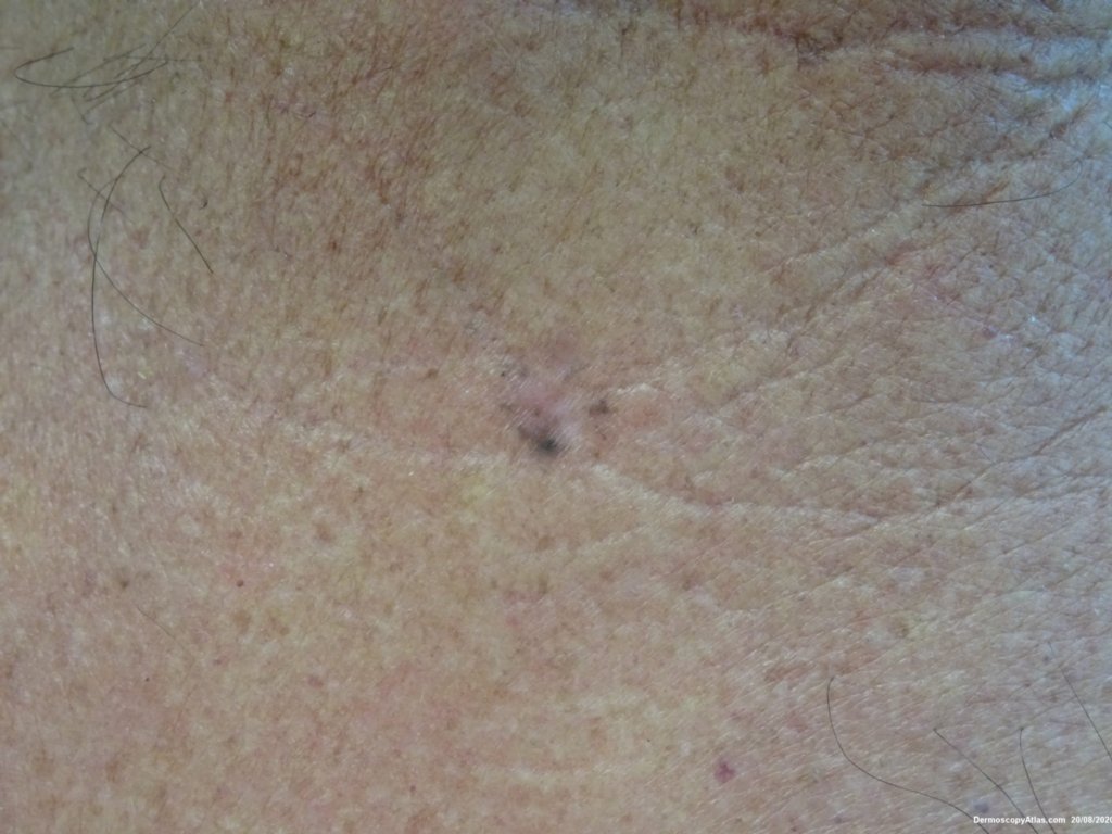
Loading
TeleDermConsult-logo
  • Recent Submissions
  • Search
  • Diagnosis
  • Quiz
  • Image comparison
  • Submit
  • Teaching Cases
  • Index
  • About

<< Back to Diagnosis Page

Image Number #3572 (Pigmented basal cell carcinoma)

Site: Neck nape

Diagnosis: Pigmented basal cell carcinoma

Sex: M

Age: 70

Type: Dermlite Polarised

Submitted By: Ian McColl

View Full Size Image

Click Here to View Details

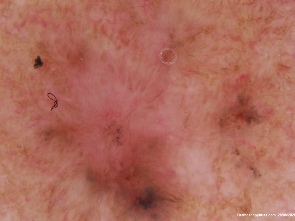
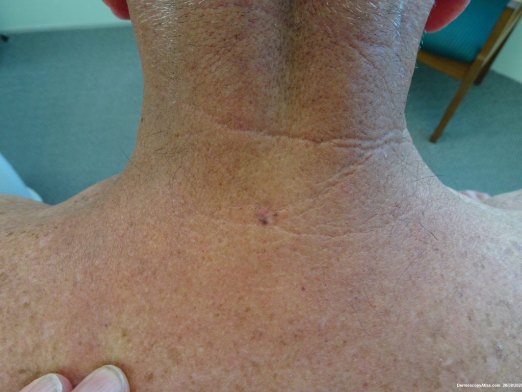
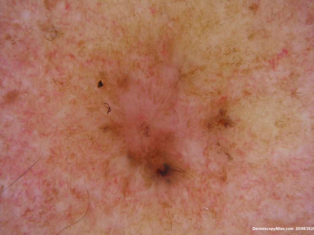
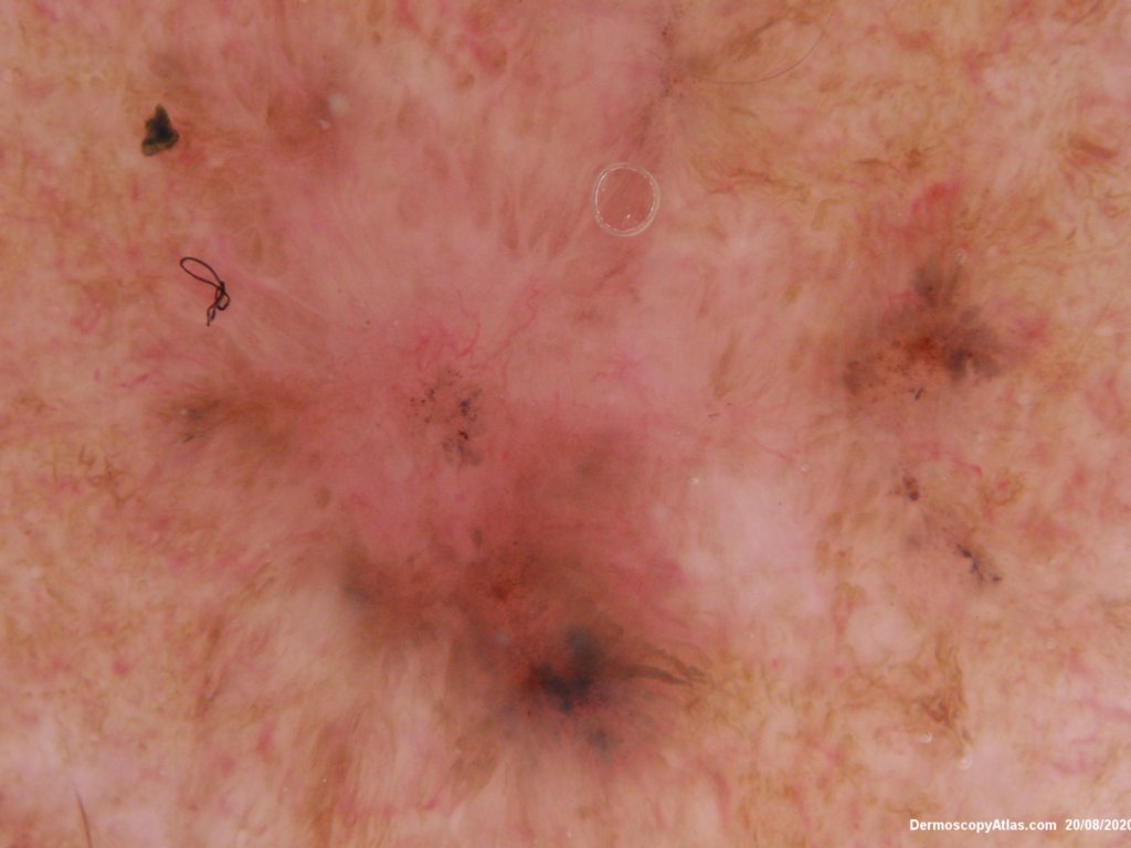
Description: Clinical dark macule neck

History:

 Clinically this lesion could easily look like a melanoma with areas of pale regression. However dermatoscopically it shows linear focussed vessels, dark blue clods and grey dots. There is no netwok. These features make it a pigmented basal cell carcinoma.

See Pigmented BCC in Dermoscopy Made Simple

DermNetNZ   eMedicine   PubMed   Dermatology Online   Archives   JAAD for "Pigmented basal cell carcinoma"

  • Recent Submissions
  • Search
  • Diagnosis
  • Quiz
  • Image comparison
  • Submit an Image
  • Teaching Cases
  • Index
  • About

Copyright 2022. Dermoscopy Atlas. ALL Rights Reserved.

Change Image