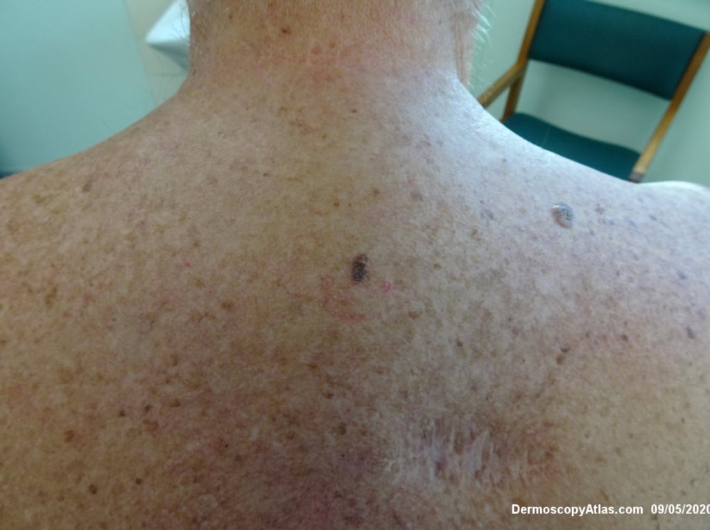
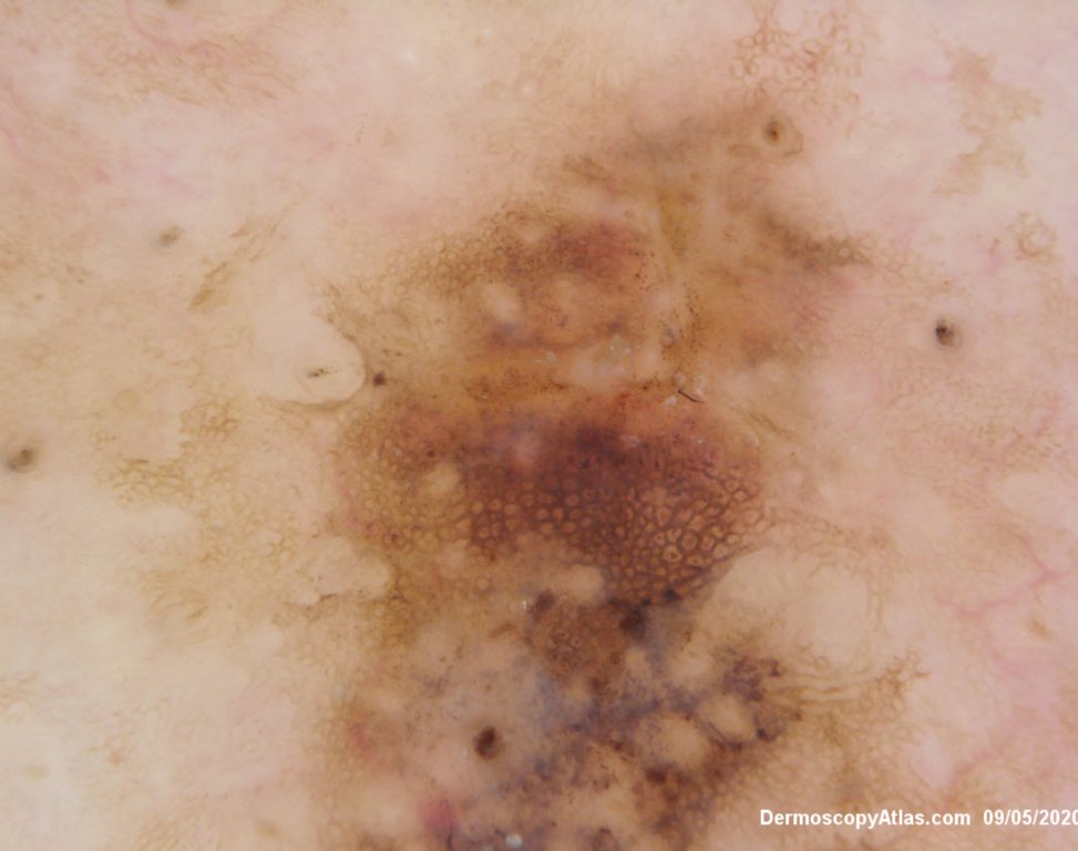
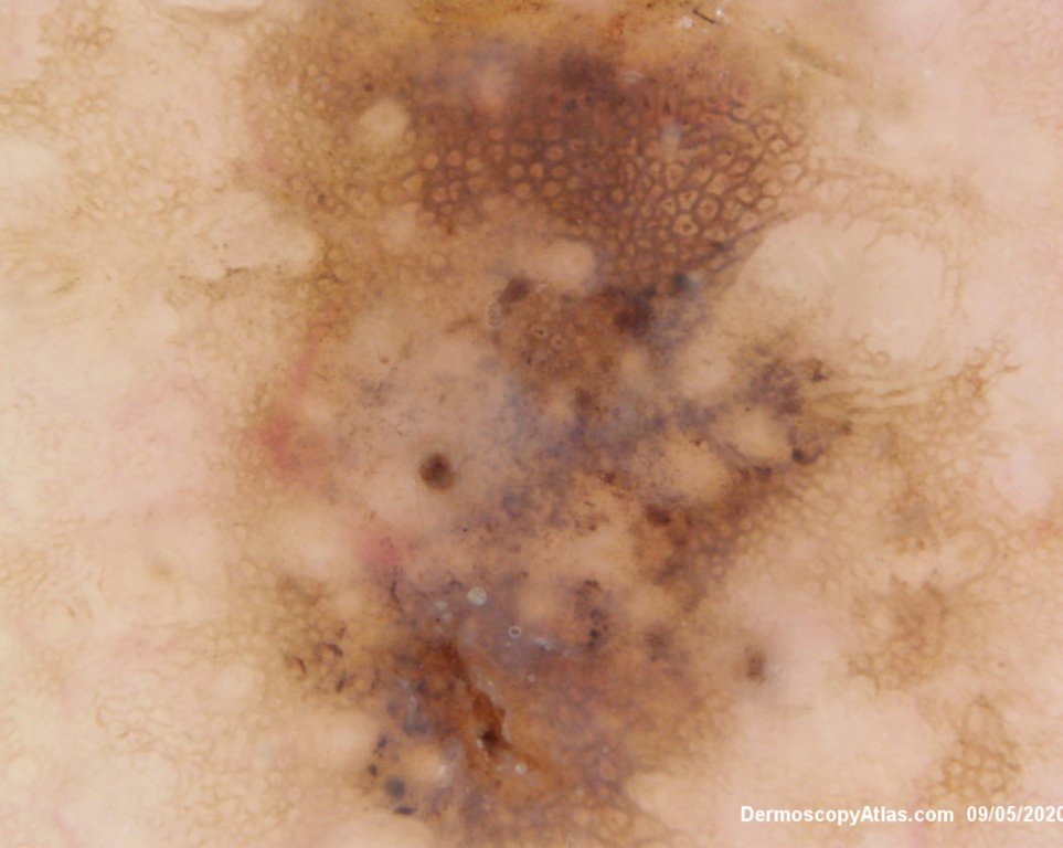
Loading
TeleDermConsult-logo
  • Recent Submissions
  • Search
  • Diagnosis
  • Quiz
  • Image comparison
  • Submit
  • Teaching Cases
  • Index
  • About

<< Back to Diagnosis Page

Image Number #3564 (Melanoma in situ)

Site: Back

Diagnosis: Melanoma in situ

Sex: M

Age: 70

Type: Dermlite Polarised

Submitted By: Ian McColl

View Full Size Image

Click Here to View Details

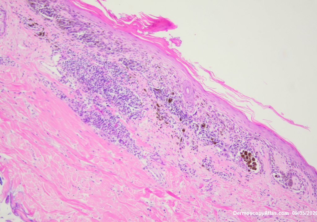
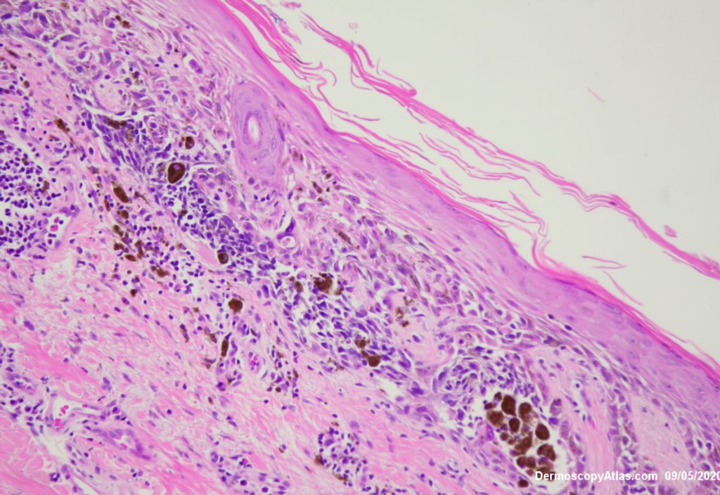
Description: Histology

History:

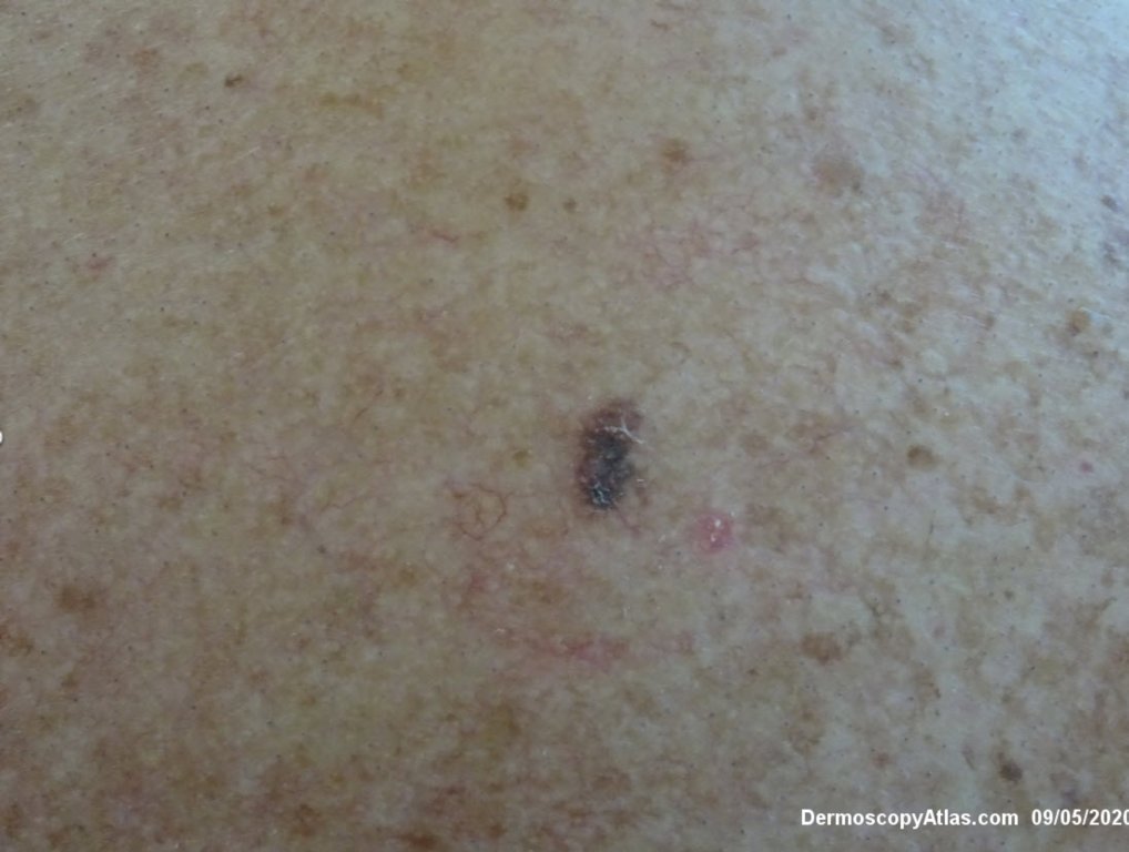
 Lesion on the back noted during a skin examination. Patient had a past history of 2 melanomas. Was this a regressing solar lentigo or a melanoma in situ?

This was a melanoma in situ with atypical melanocytes in nests and along the Dermoepidermal junction.

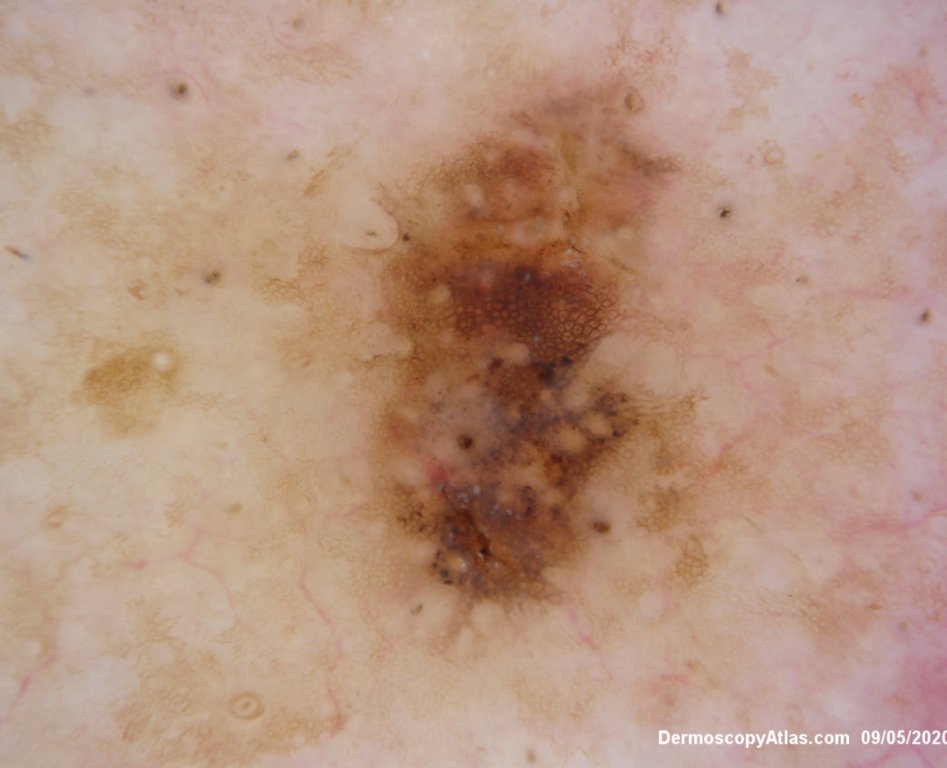
Dermatoscopically there was a thickened network with grey dots of regression and polygon formation.

Below is a video on solar lentigo for comparison.


DermNetNZ   eMedicine   PubMed   Dermatology Online   Archives   JAAD for "Melanoma in situ"

  • Recent Submissions
  • Search
  • Diagnosis
  • Quiz
  • Image comparison
  • Submit an Image
  • Teaching Cases
  • Index
  • About

Copyright 2022. Dermoscopy Atlas. ALL Rights Reserved.

Change Image