
Site: Back
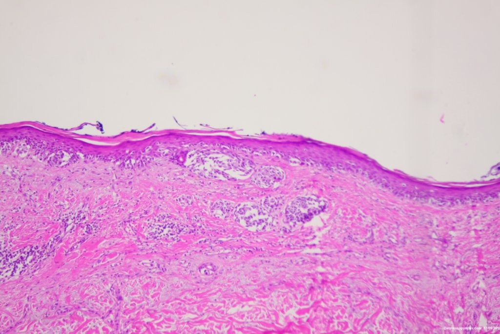
Diagnosis: Melanoma regression
Sex: M
Age: 68
Type: Dermlite Polarised
Submitted By: Ian McColl
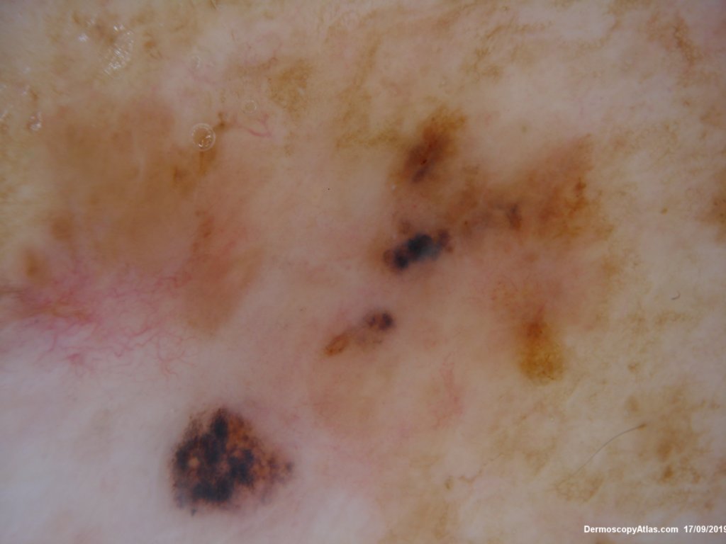
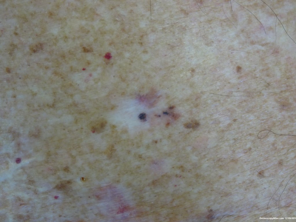
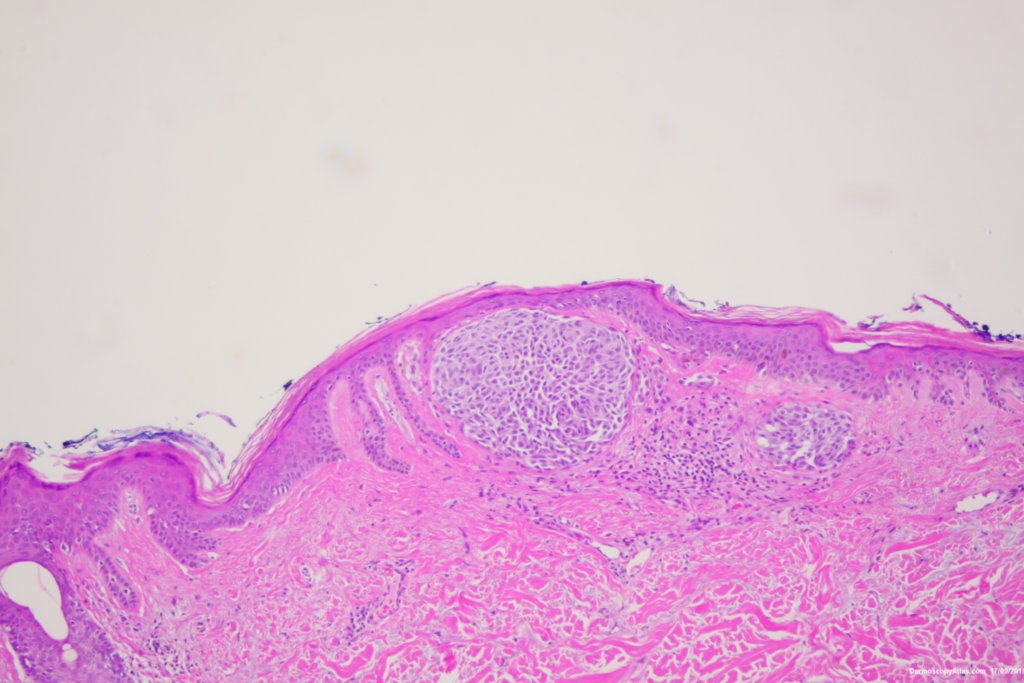
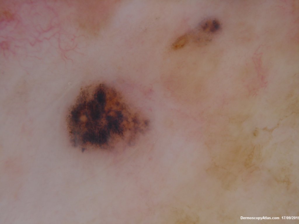
Description: Pigmentation in regressed area
History:
Clinically the lesion looked like a pigmented BCC but the dermatoscopy was equivocal with rtegression, blue black clods and some black dots. It was an invasive melanoma 0.38 mm thick arising in a lentigo maligna with regression changes.