
Site: Shoulder
Diagnosis: Melanoma in situ
Sex: F
Age: 65
Type: Heine
Submitted By: Ian McColl
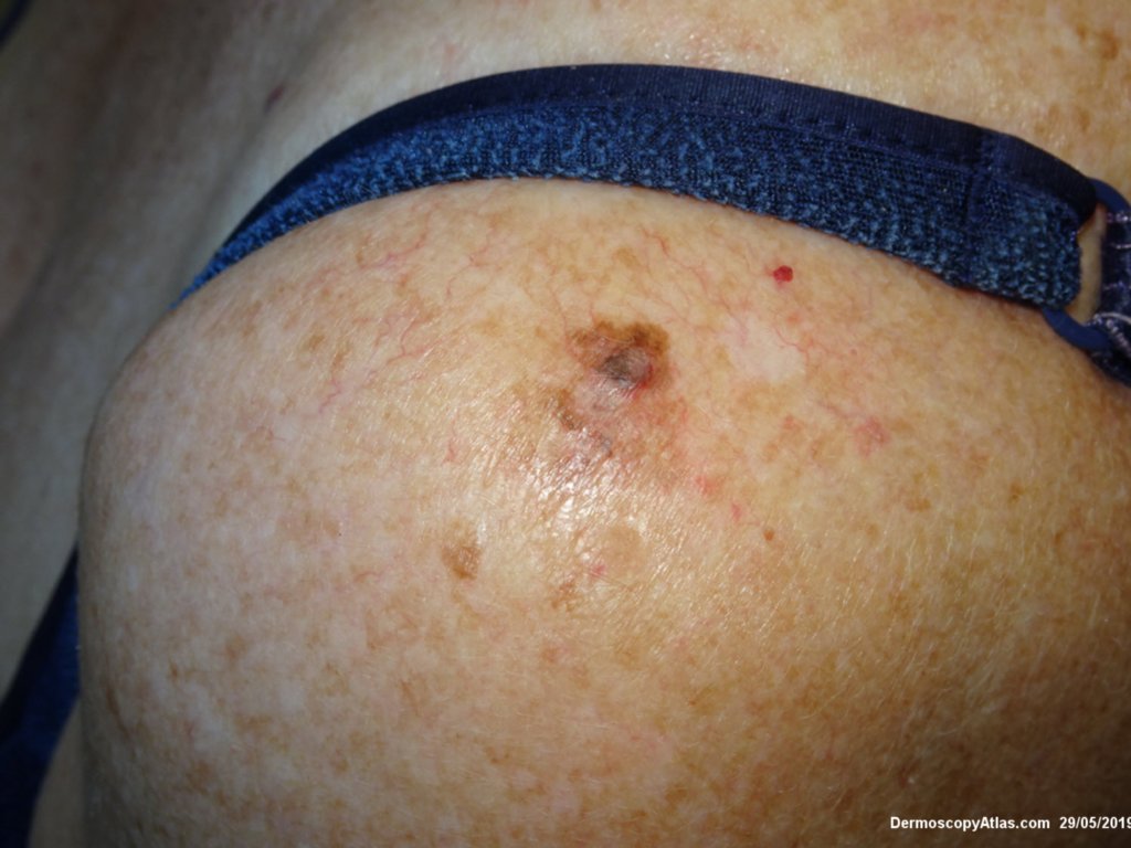
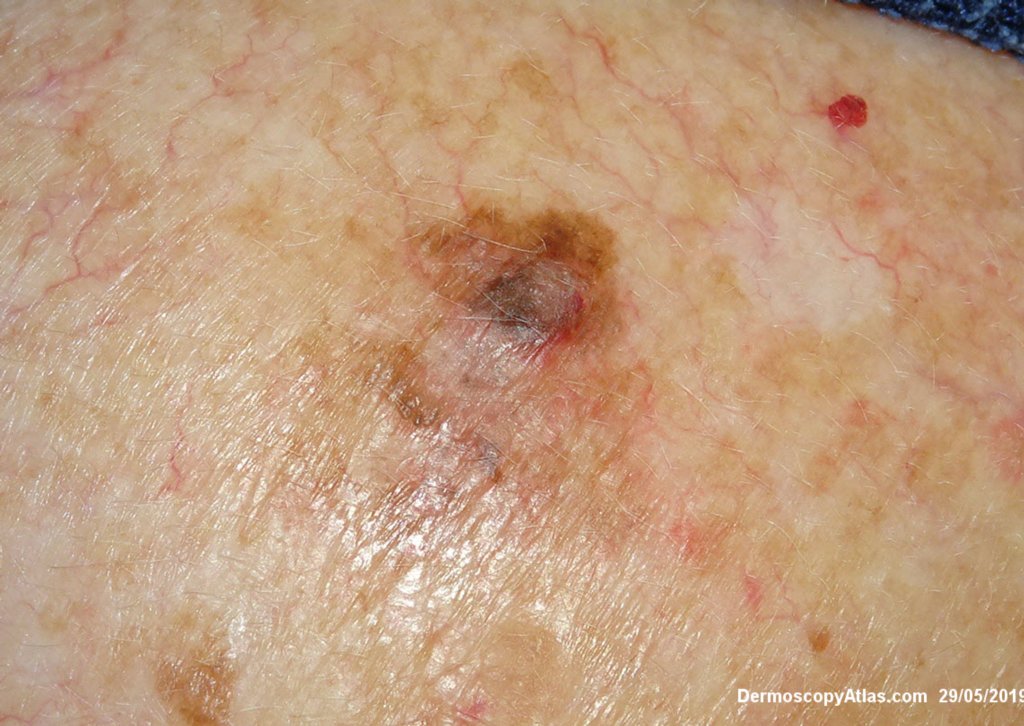
Description: Clinical close up
History:
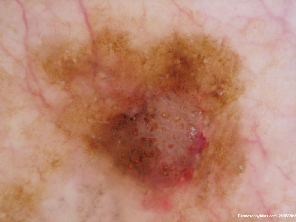
A lesion on the shoulder slowly changing. There are varying colours and the lesion seems to have a seborrhoeic keratosis at one pole.
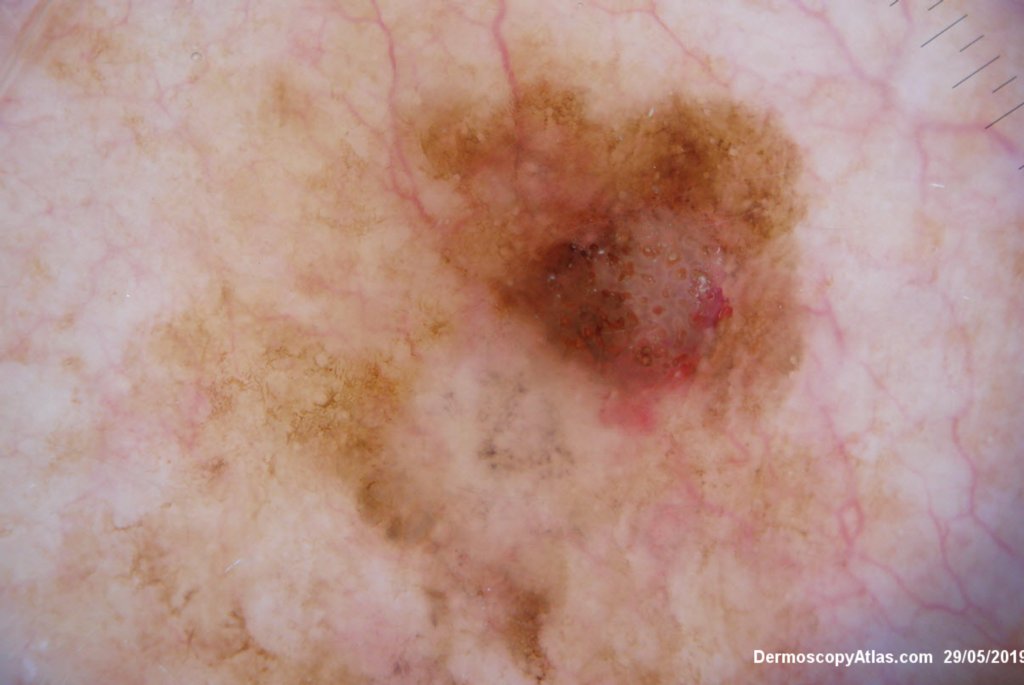
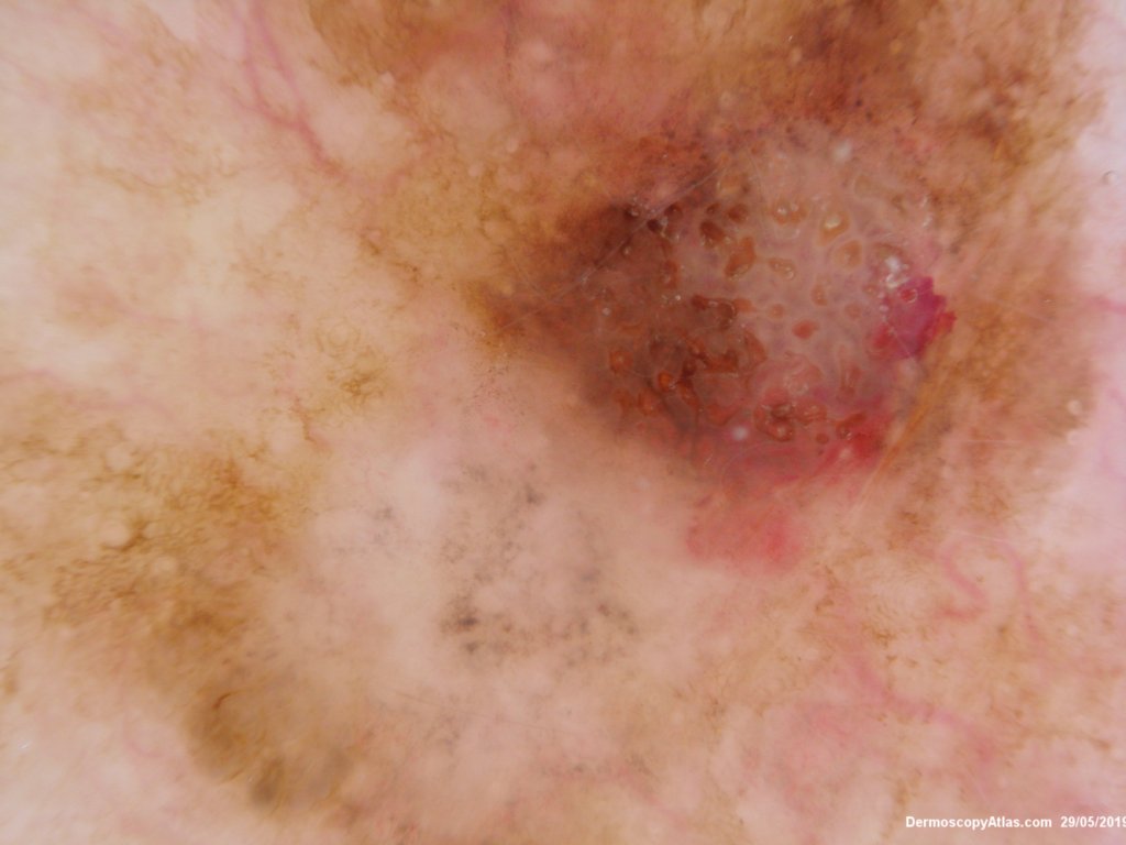
The dermatoscopy shows grey dots of regression and the orange clods in the seb K but there is also some hemorrhage within the Seb k and pigment occluding part of it.
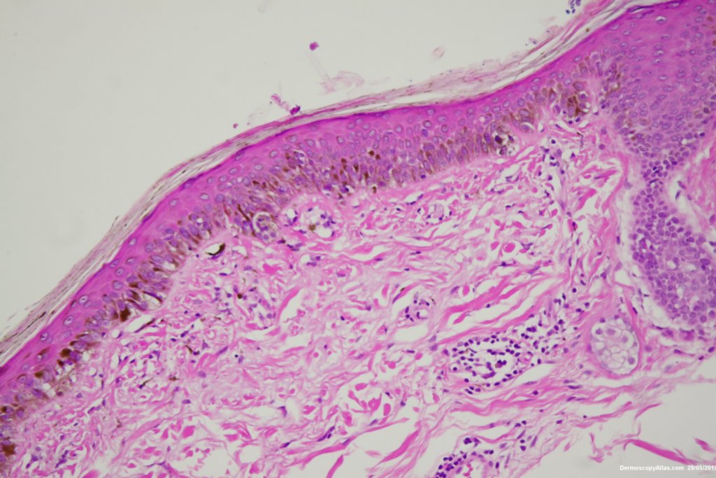
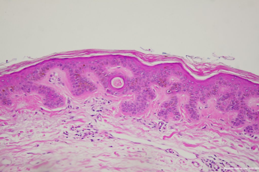
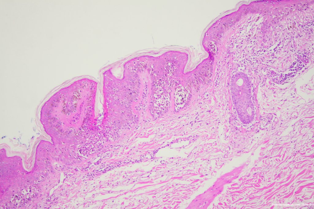
A shave biopsy removing the lesion was done. The histology showed a lentiginous proliferation of atypical basal melanocytes with a lymphocytic infiltrate and some melanin in melanophages causing the grey dots. There was also an in situ melanoma involving part of the seborrhoeic keratosis.
See a more dertailed view of the histology as the 3rd case in the video below.