
Site: Leg
Diagnosis: Melanoma invasive
Sex: M
Age: 67
Type: Dermlite Polarised
Submitted By: Ian McColl
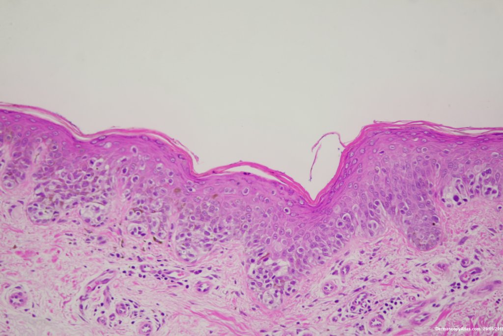
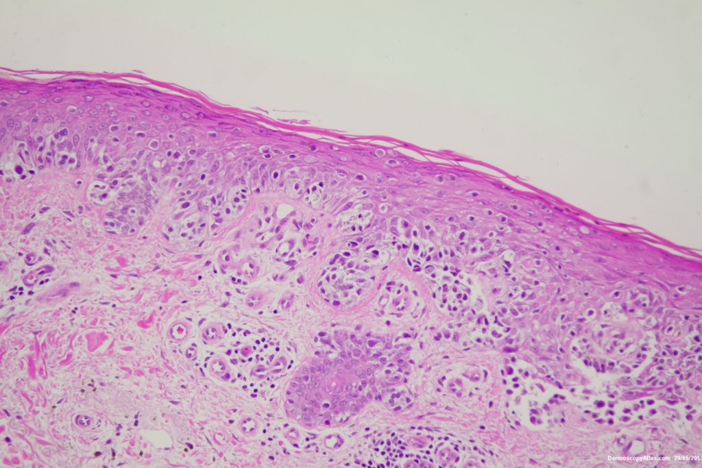
Description: Histology Mainly in situ with Pagetoid spread
History:
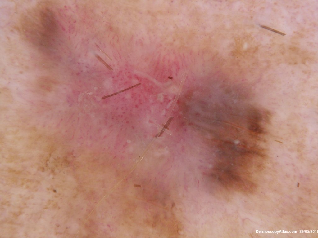
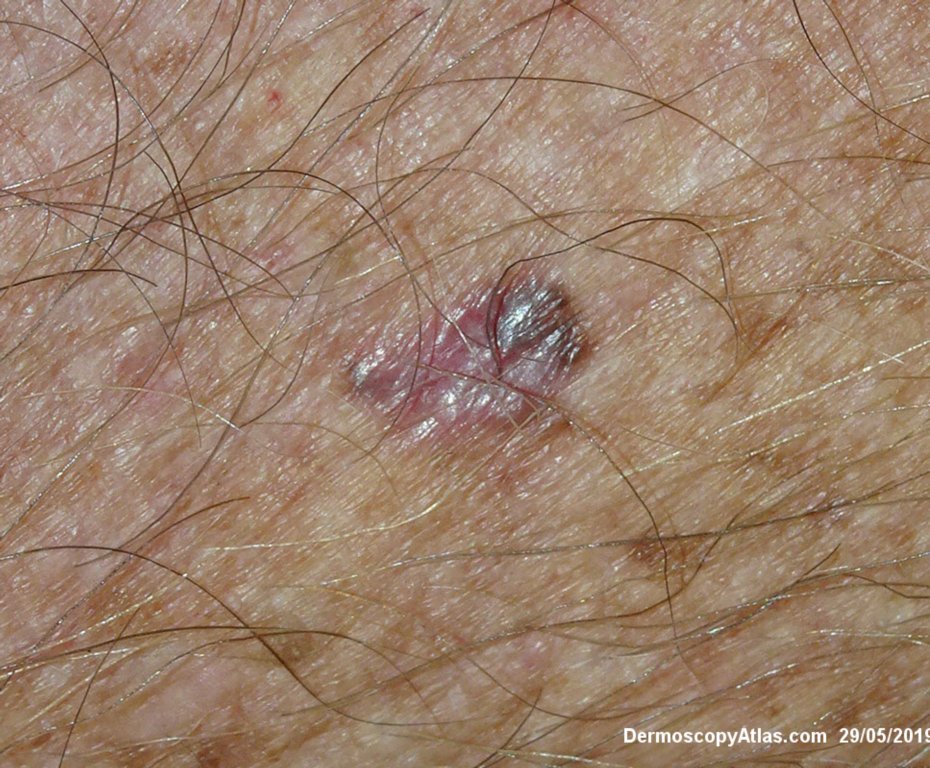
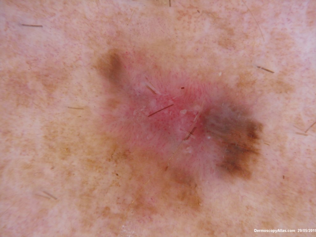
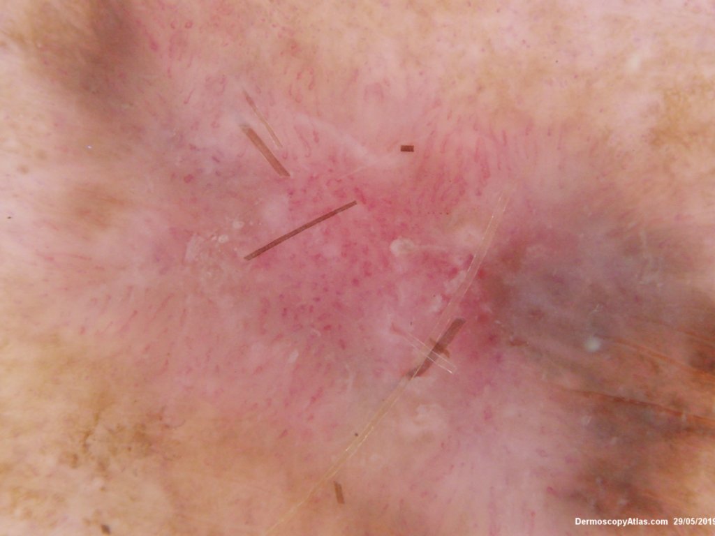
Pigmented lesion on the lower leg noted at an annual skin check. The clinical is suggestive of a melanoma but the dermatoscopy was initially interpreted as coiled vessels with lines radial peripheral of an IEC. In IEC the lines radial are usually made up of dots but these are solid streaks. There is some vessel variability in the pink area.
This was an invasive melanoma 0.4 mm thick but the histology shown shows mainly in situ melanoma.Have a look at the second case in the video below for a more detailed overview of the histology.