Loading
TeleDermConsult-logo
  • Recent Submissions
  • Search
  • Diagnosis
  • Quiz
  • Image comparison
  • Submit
  • Teaching Cases
  • Index
  • About

<< Back to Diagnosis Page

Image Number #3468 (Melanoma in situ)

Site: Abdomen

Diagnosis: Melanoma in situ

Sex: M

Age: 67

Type: Dermlite Polarised

Submitted By: Ian McColl

View Full Size Image

Click Here to View Details

Description: Histology

History:

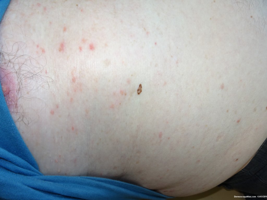
 This lesion was noted on the abdominal wall during a routine skin examination. Again the patient had not been aware of it. 

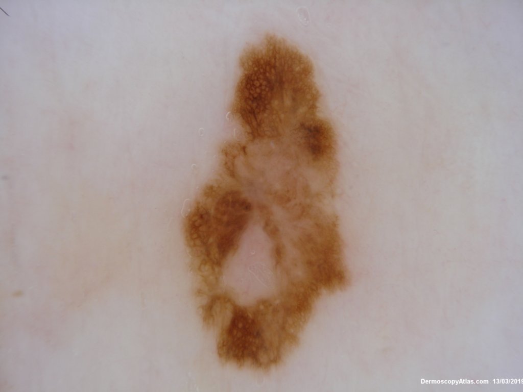
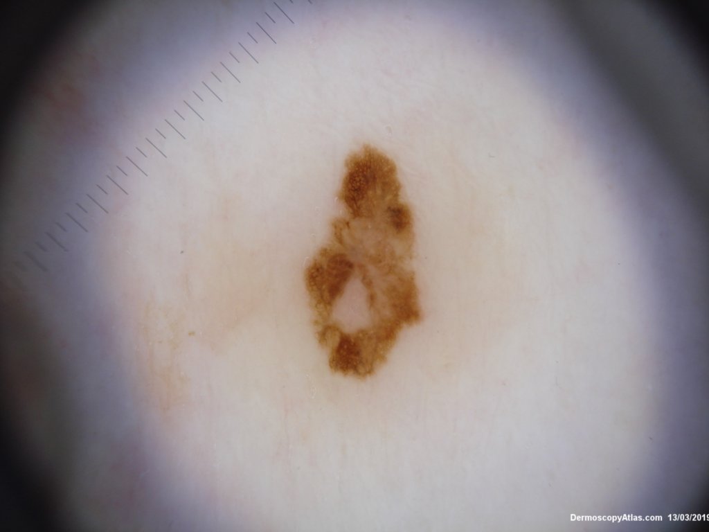
Dermatoscopy showed a thickened network in areas with some loss centrally. There were a few radial segmental lines and a suggestion of grey. 

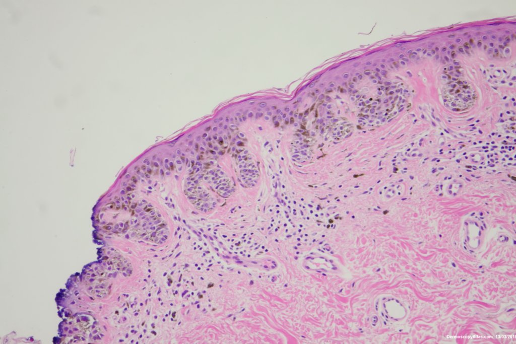
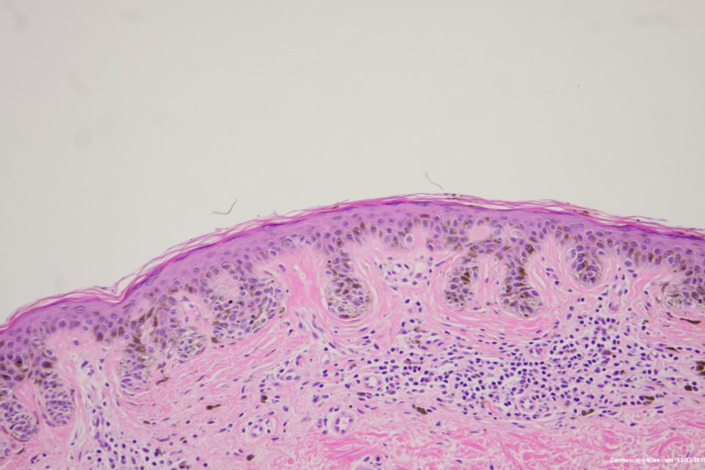
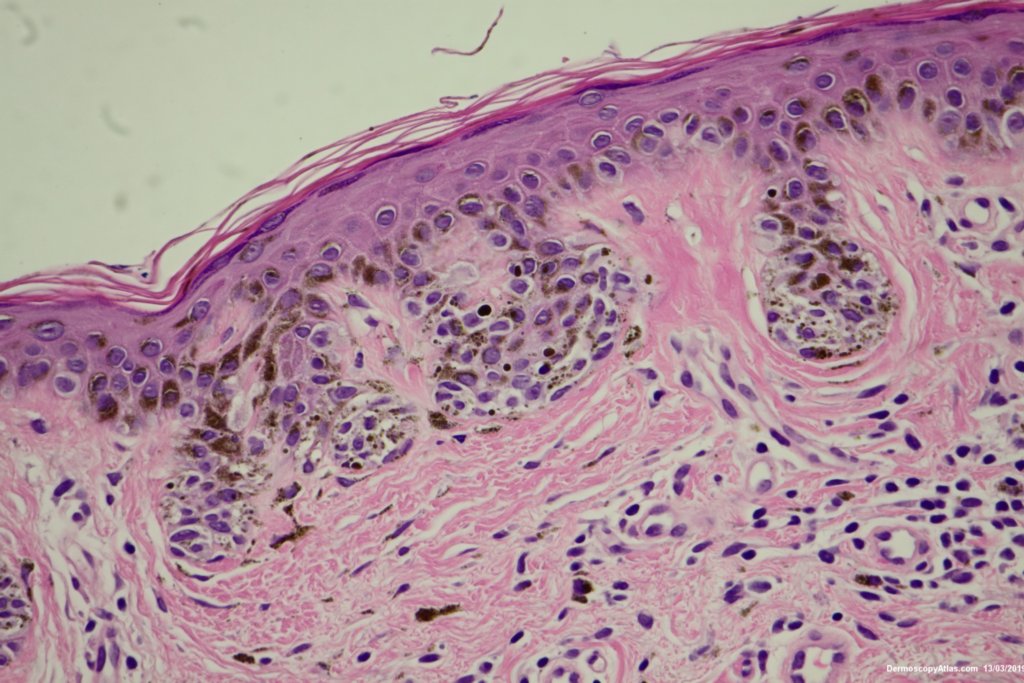
Histology showed an in situ melanoma with nests of atypical melanocytes in the rete ridges. No real pagetoid spread was seen.

Excision with 5 mm clinical margins after the initial shave biopsy.

DermNetNZ   eMedicine   PubMed   Dermatology Online   Archives   JAAD for "Melanoma in situ"

  • Recent Submissions
  • Search
  • Diagnosis
  • Quiz
  • Image comparison
  • Submit an Image
  • Teaching Cases
  • Index
  • About

Copyright 2022. Dermoscopy Atlas. ALL Rights Reserved.

Change Image