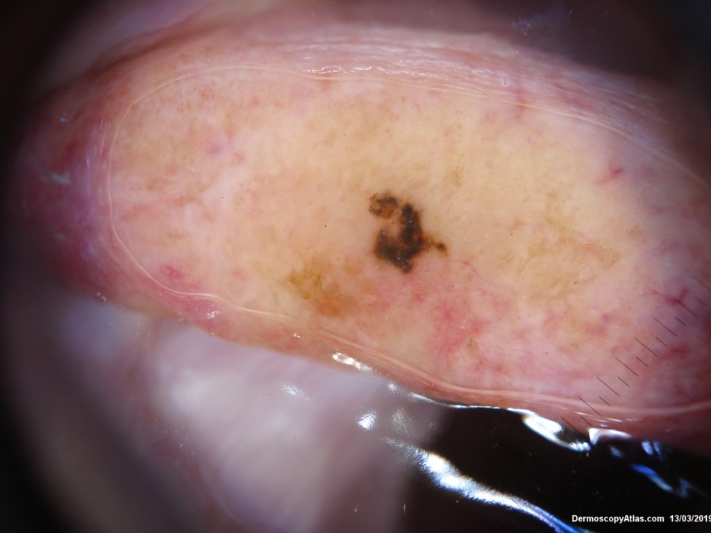
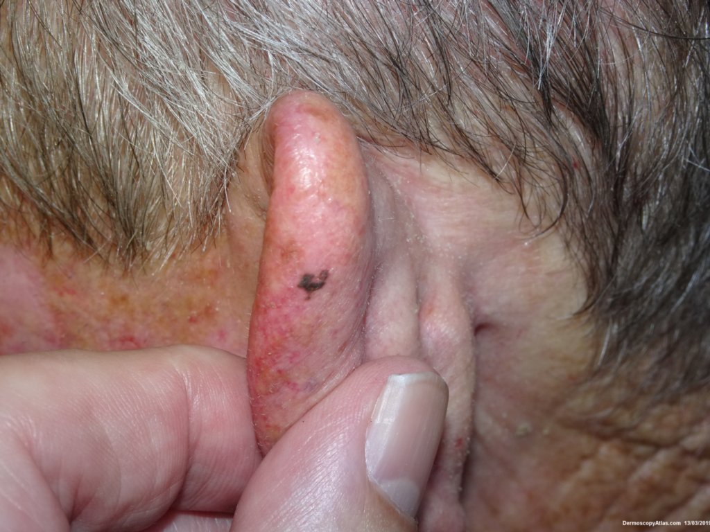
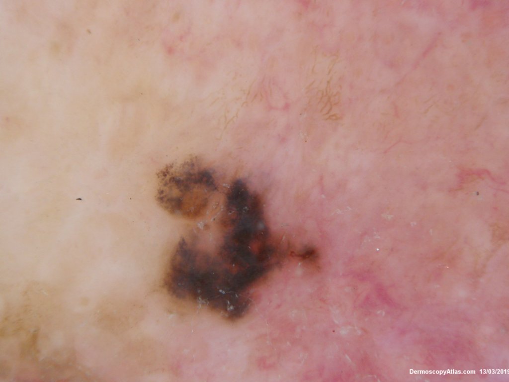
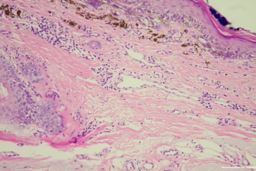
Loading
TeleDermConsult-logo
  • Recent Submissions
  • Search
  • Diagnosis
  • Quiz
  • Image comparison
  • Submit
  • Teaching Cases
  • Index
  • About

<< Back to Diagnosis Page

Image Number #3456 (Melanoma in situ)

Site: Ear

Diagnosis: Melanoma in situ

Sex: M

Age: 70

Type: Dermlite Polarised

Submitted By: Ian McColl

View Full Size Image

Click Here to View Details

Description: Dermatoscopy

History:

 This lesion was noted on the ear during a routine skin check. Patient was not aware of it. The dermatoscopy showed an irregular blue black structureless lesion with some peripheral grey dots as peppering, an excentric area of regression and some white clods.

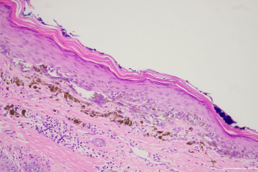
The histology was reported as an in situ melanoma with some dermal regression. It was excised with 5 mm clinical margins after the initial narrow excision biopsy.

DermNetNZ   eMedicine   PubMed   Dermatology Online   Archives   JAAD for "Melanoma in situ"

  • Recent Submissions
  • Search
  • Diagnosis
  • Quiz
  • Image comparison
  • Submit an Image
  • Teaching Cases
  • Index
  • About

Copyright 2022. Dermoscopy Atlas. ALL Rights Reserved.

Change Image