
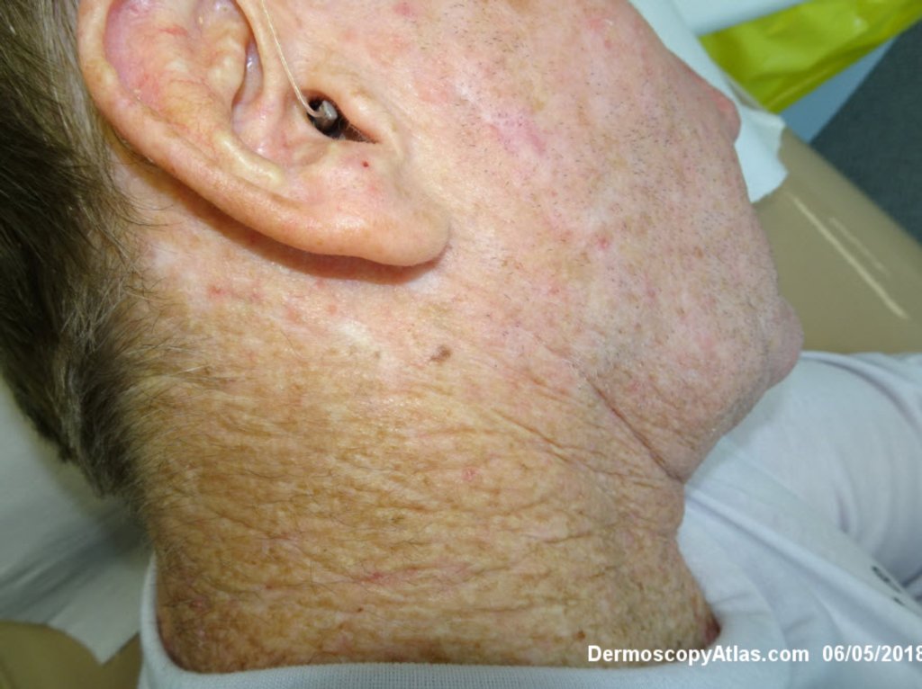
Site: Neck side
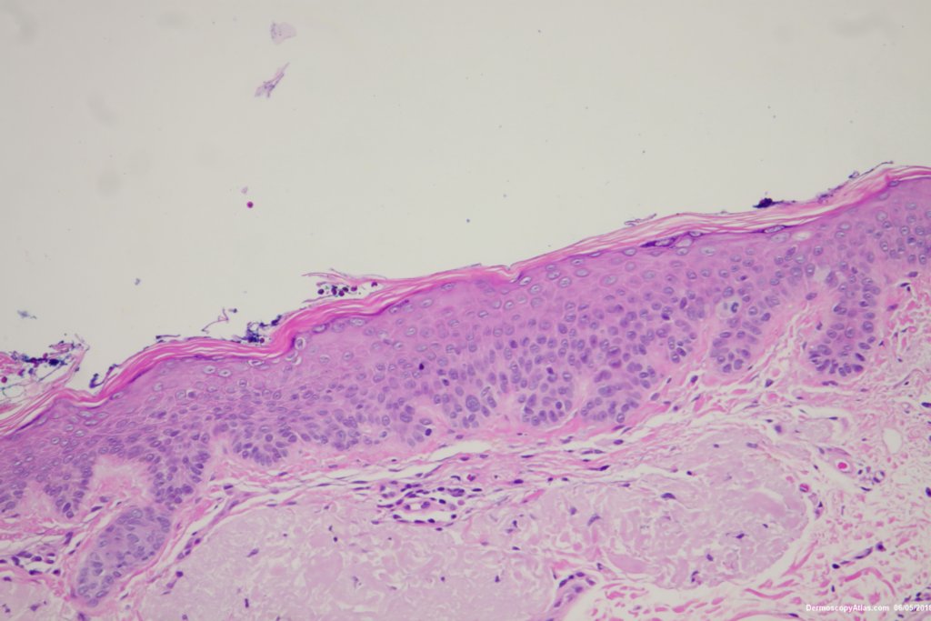
Diagnosis: Pigmented Intraepidermal carcinoma
Sex: M
Age: 67
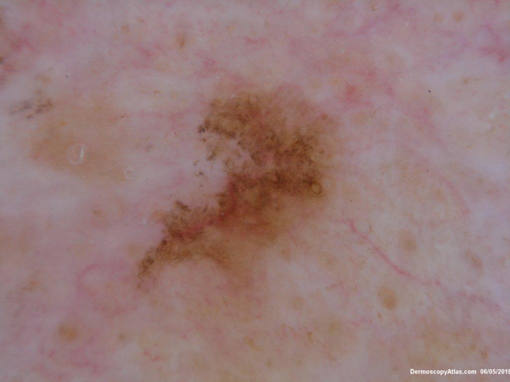
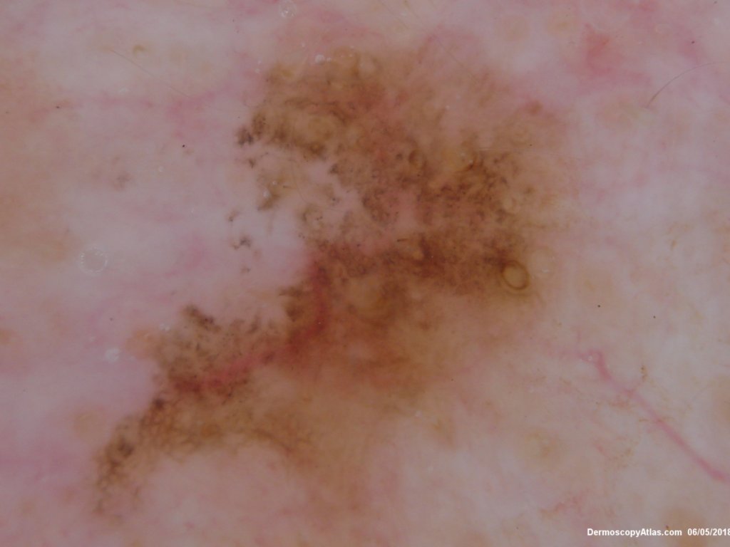
Type: Dermlite Polarised
Submitted By: Ian McColl
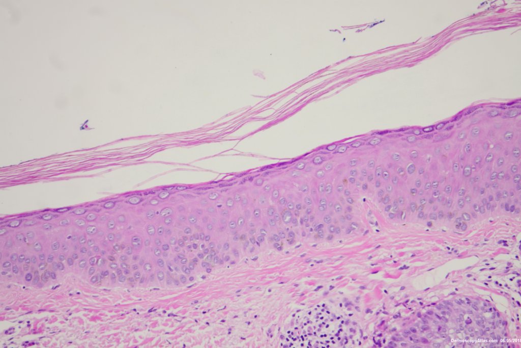
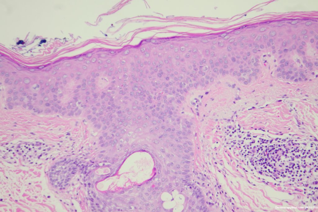
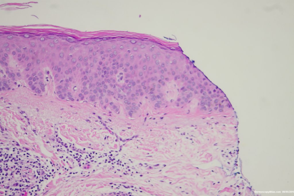
Description: Histology Pigmented IEC
History:
Some pigmented lesions look melanocytic, However this is a pigmented intraepidermal carcinoma. Some areas show more full thickness atypia than others. The dermatoscopy shows some dots in rows but there are other grey dots showing regression at one edge.