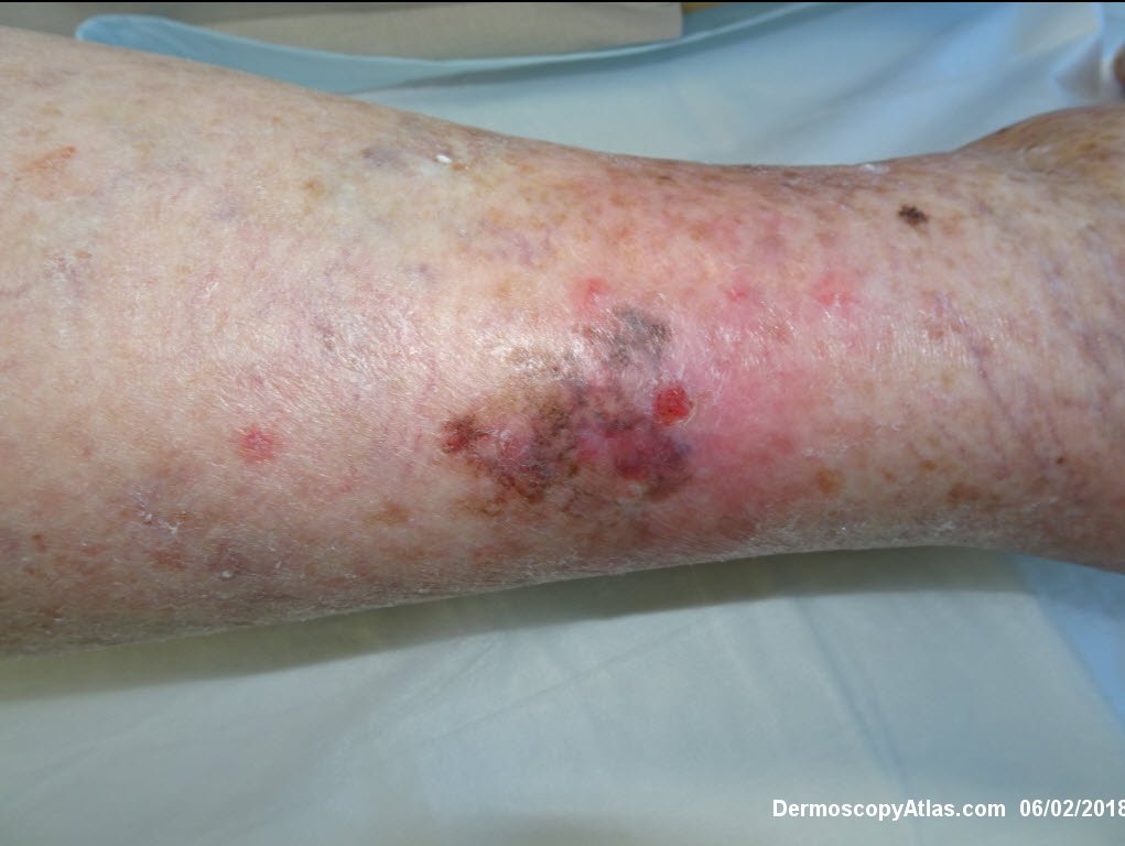
Loading
TeleDermConsult-logo
  • Recent Submissions
  • Search
  • Diagnosis
  • Quiz
  • Image comparison
  • Submit
  • Teaching Cases
  • Index
  • About

<< Back to Diagnosis Page

Image Number #3421 (Melanoma invasive)

Site: Leg

Diagnosis: Melanoma invasive

Sex: F

Age: 84

Type: Dermlite Polarised

Submitted By: Ian McColl

View Full Size Image

Click Here to View Details

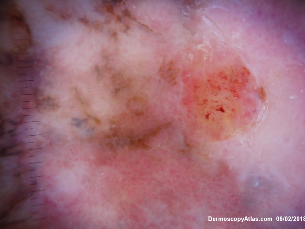
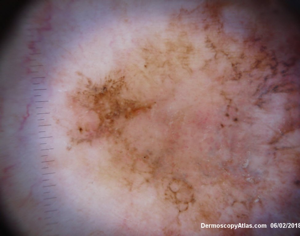
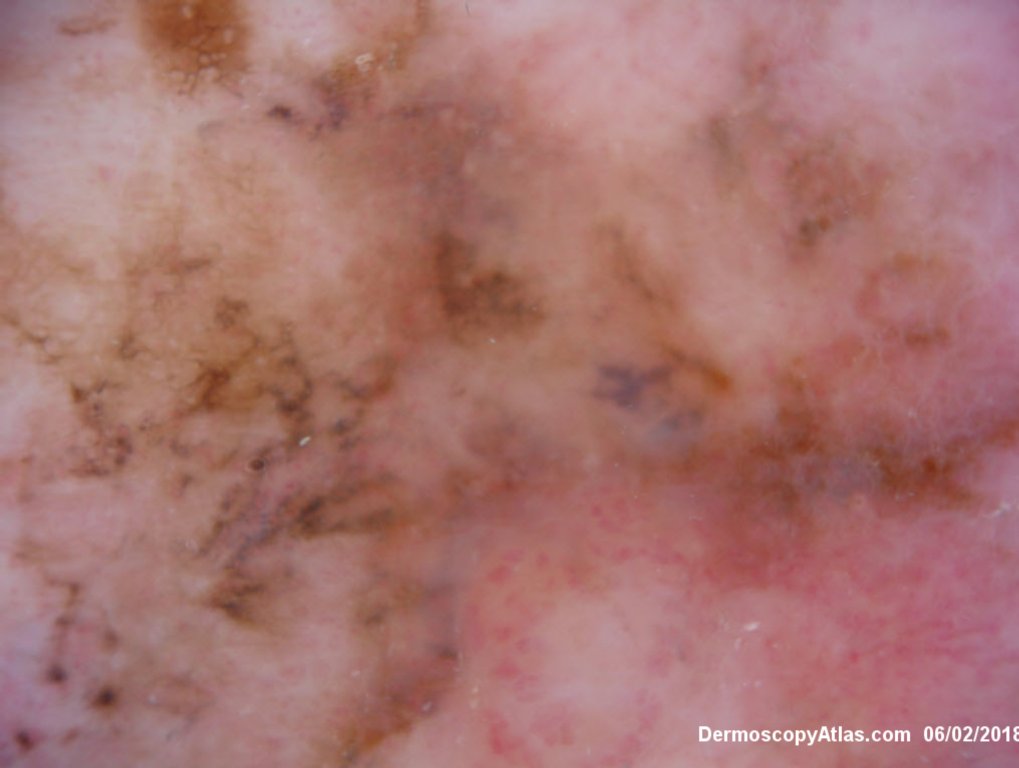
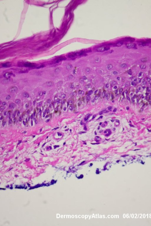
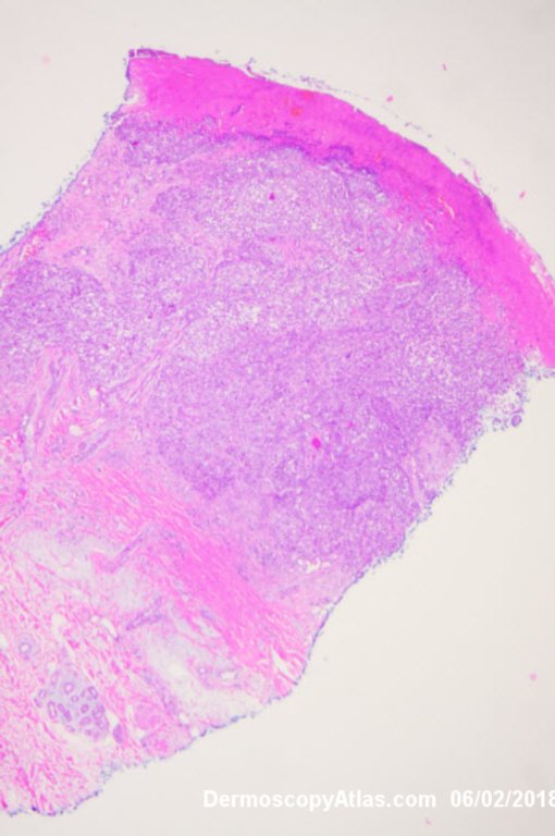
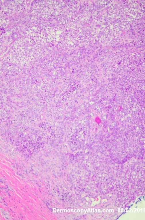
Description: Multiple colours in this pigmented lesion on the lower leg. New pink area

History:

   Lady in her early 80s who presented with this lesion on her ankle. The pigmented area had been there for some time but the pink area was bleeding and new. Shave of surrounding area and a punch biopsy of the new pink area showed mainly surrounding in situ melanoma with invasive melanoma in the pink area 1.8 mm thick. Having a 2cms margin excision of the whole area and a graft.

DermNetNZ   eMedicine   PubMed   Dermatology Online   Archives   JAAD for "Melanoma invasive"

  • Recent Submissions
  • Search
  • Diagnosis
  • Quiz
  • Image comparison
  • Submit an Image
  • Teaching Cases
  • Index
  • About

Copyright 2022. Dermoscopy Atlas. ALL Rights Reserved.

Change Image