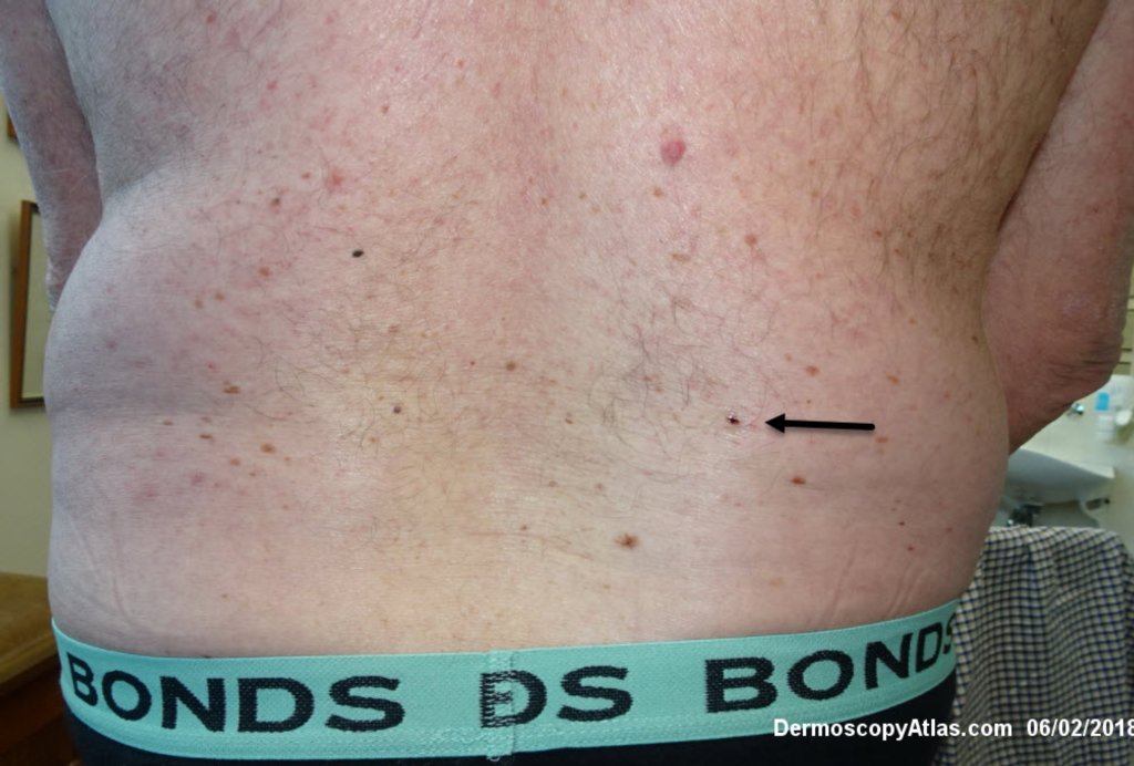
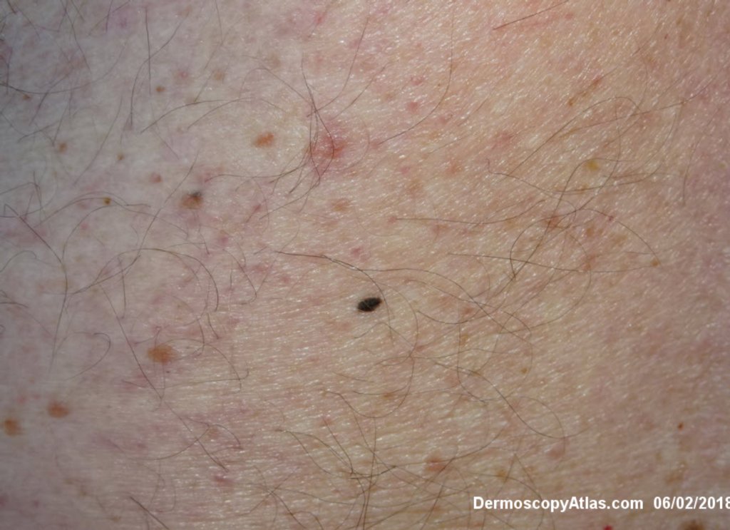
Loading
TeleDermConsult-logo
  • Recent Submissions
  • Search
  • Diagnosis
  • Quiz
  • Image comparison
  • Submit
  • Teaching Cases
  • Index
  • About

<< Back to Diagnosis Page

Image Number #3418 (Melanoma invasive)

Site: Back

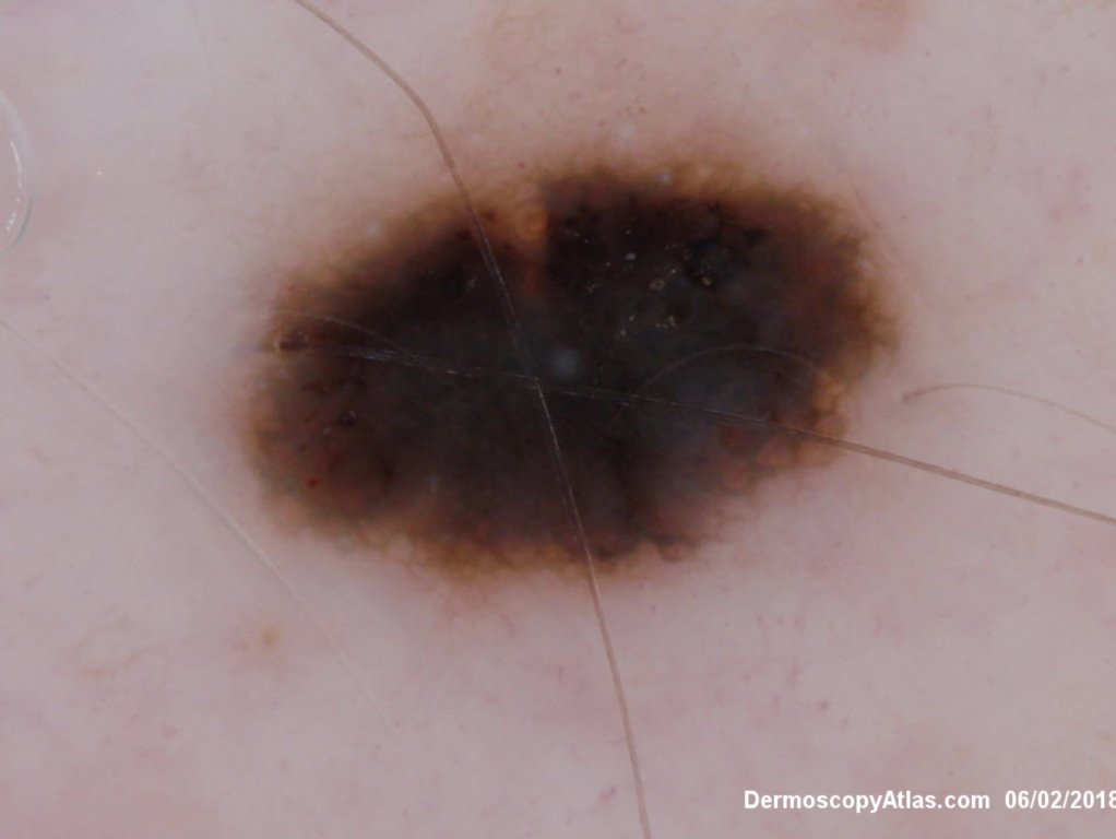
Diagnosis: Melanoma invasive

Sex: M

Age: 67

Type: Dermlite Polarised

Submitted By: Ian McColl

View Full Size Image

Click Here to View Details

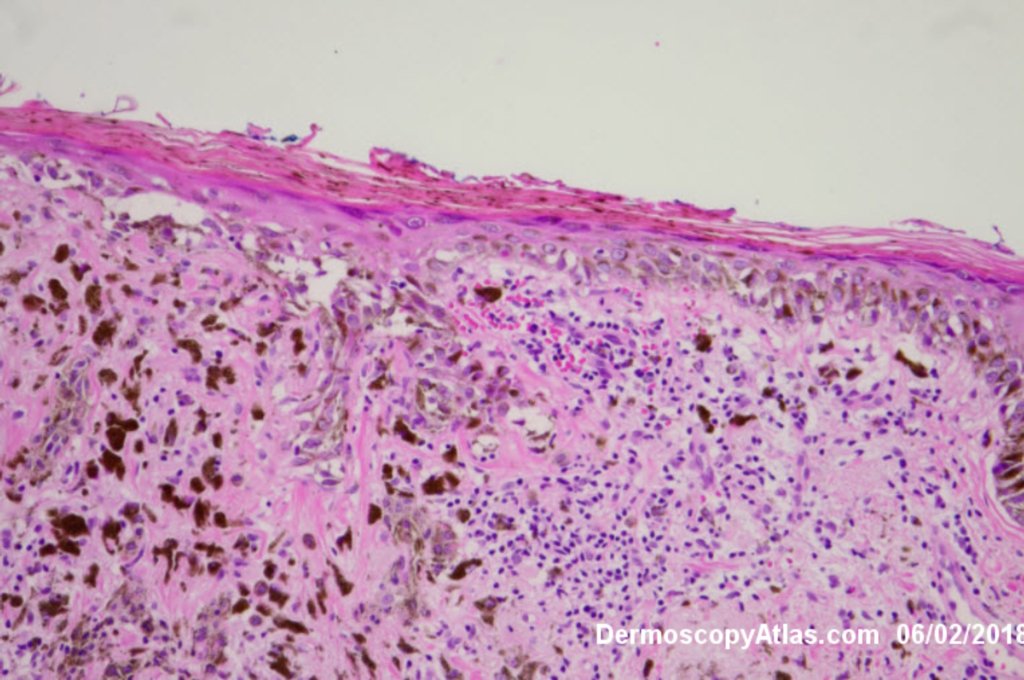
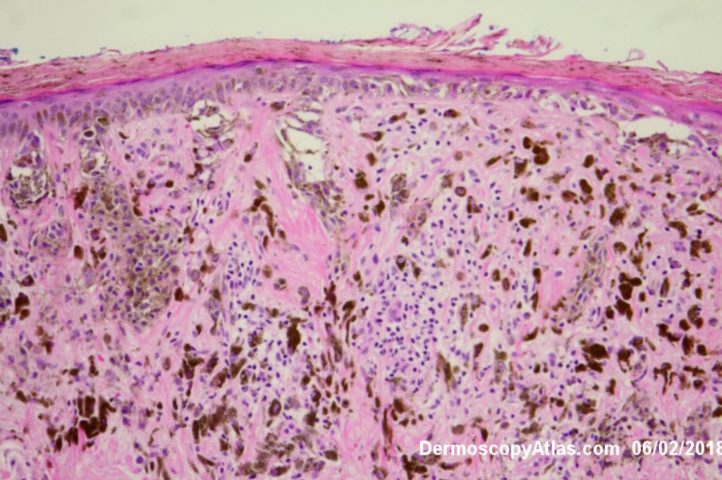
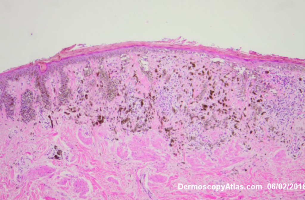
Description: Histology showing in situ melanoma and dermal pigmentation

History:

 Routine skin check of a male in late 60s with this pigmented lesion on the lower back. Not aware of it. Clinically it stood out because it was much darker than his other nevi.

Histology showed mainly an in situ melanoma with an invasive component of 0.4 mm and a lot of dermal melanin in melanophages

DermNetNZ   eMedicine   PubMed   Dermatology Online   Archives   JAAD for "Melanoma invasive"

  • Recent Submissions
  • Search
  • Diagnosis
  • Quiz
  • Image comparison
  • Submit an Image
  • Teaching Cases
  • Index
  • About

Copyright 2022. Dermoscopy Atlas. ALL Rights Reserved.

Change Image