
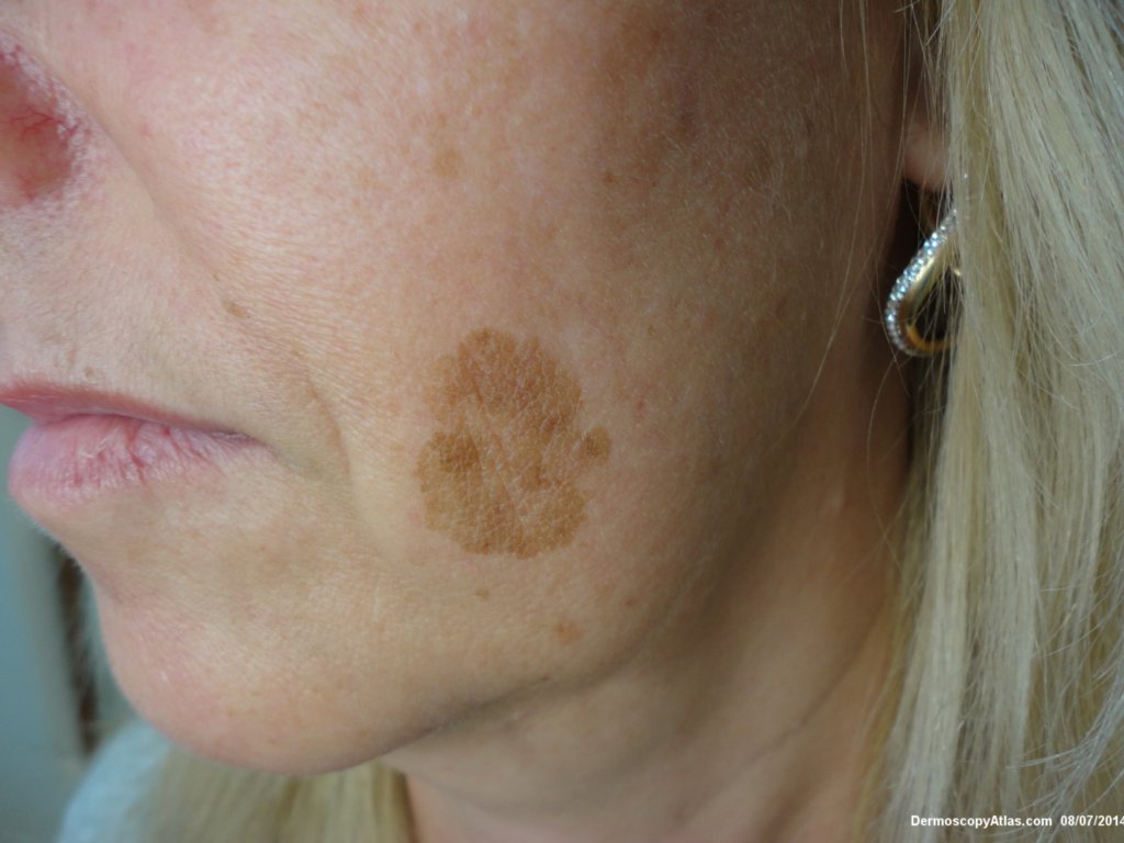
Site: Cheek
Diagnosis: Lentigene
Sex: F
Age: 44
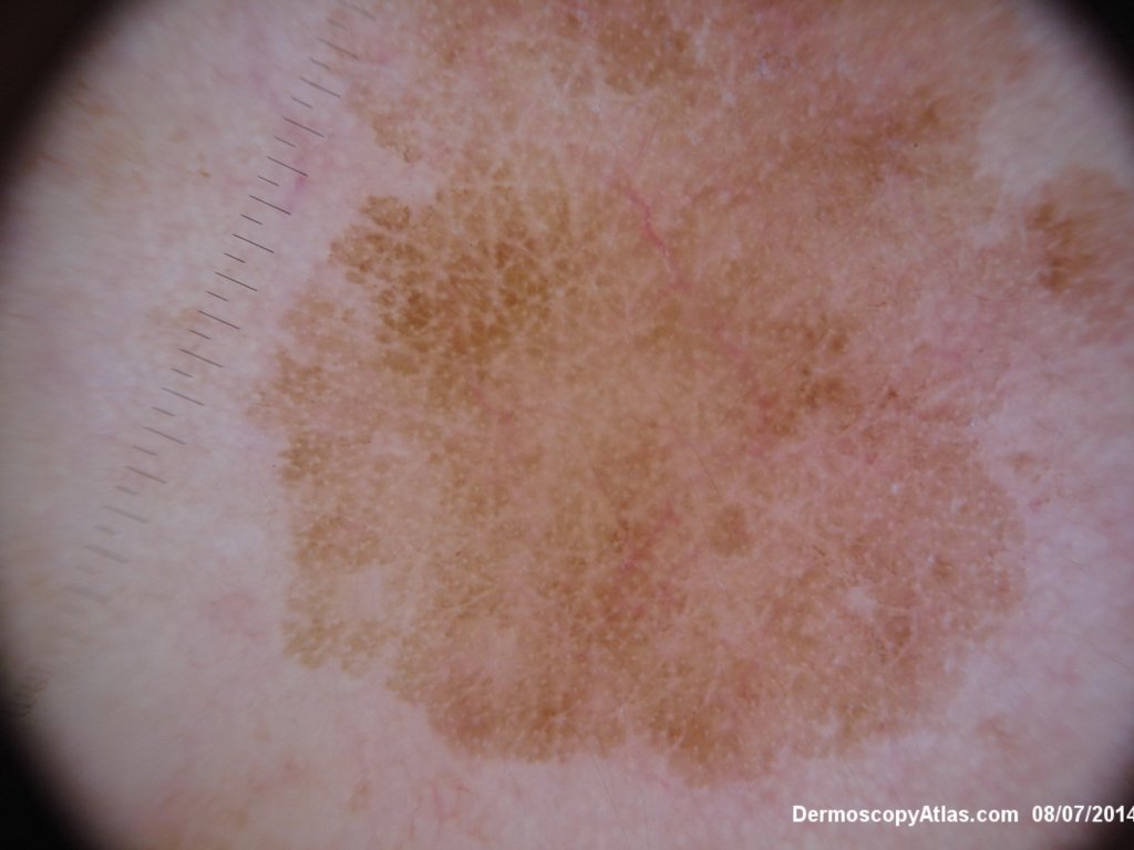
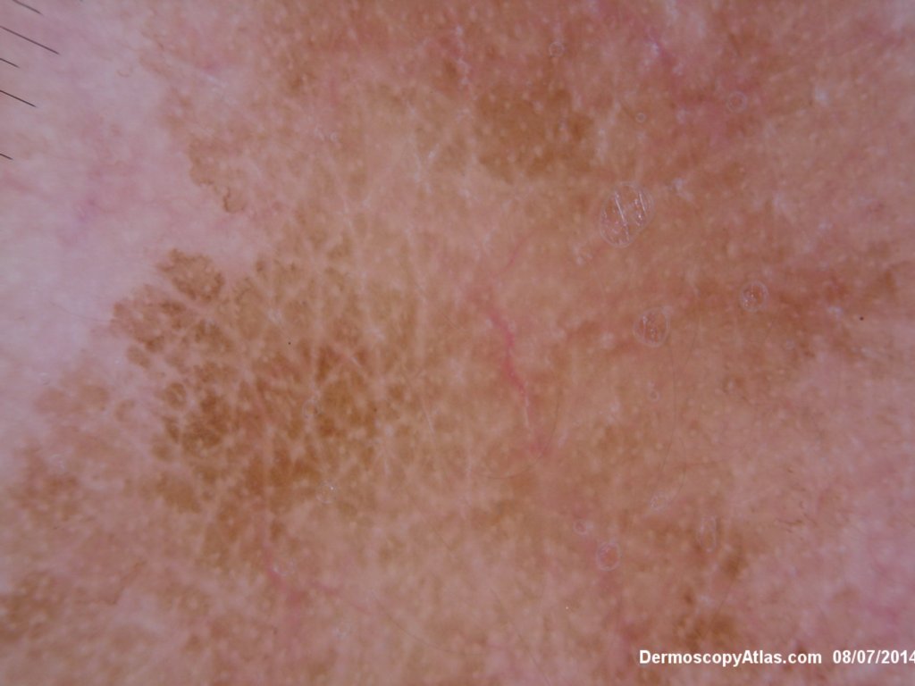
Type: Dermlite Polarised
Submitted By: Ian McColl
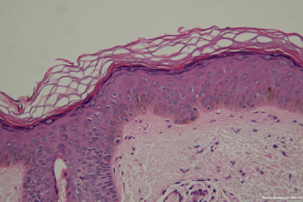
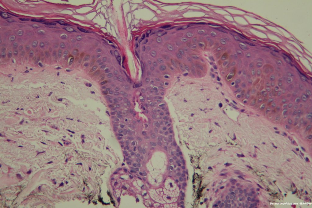
Description: Histology No evidence of excess melanocytes
History:
This lady had a slowly growing pigmented lesion on her cheek . The clour was varying a bit. The dermatoscopy did not show grey circles or other features of early lentigo maligna. Her shave biopsy showed only increased melanin in basal keratinocytes. This was a lentigene. It was lasered off for cosmetic reasons.