
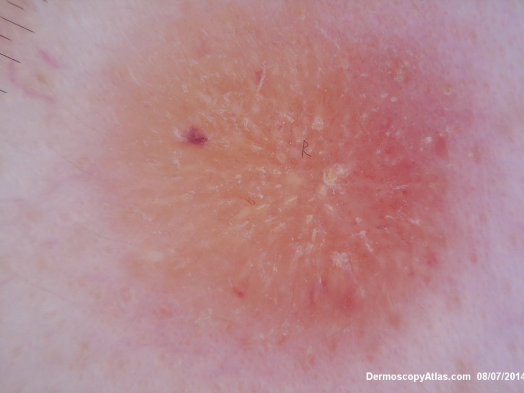
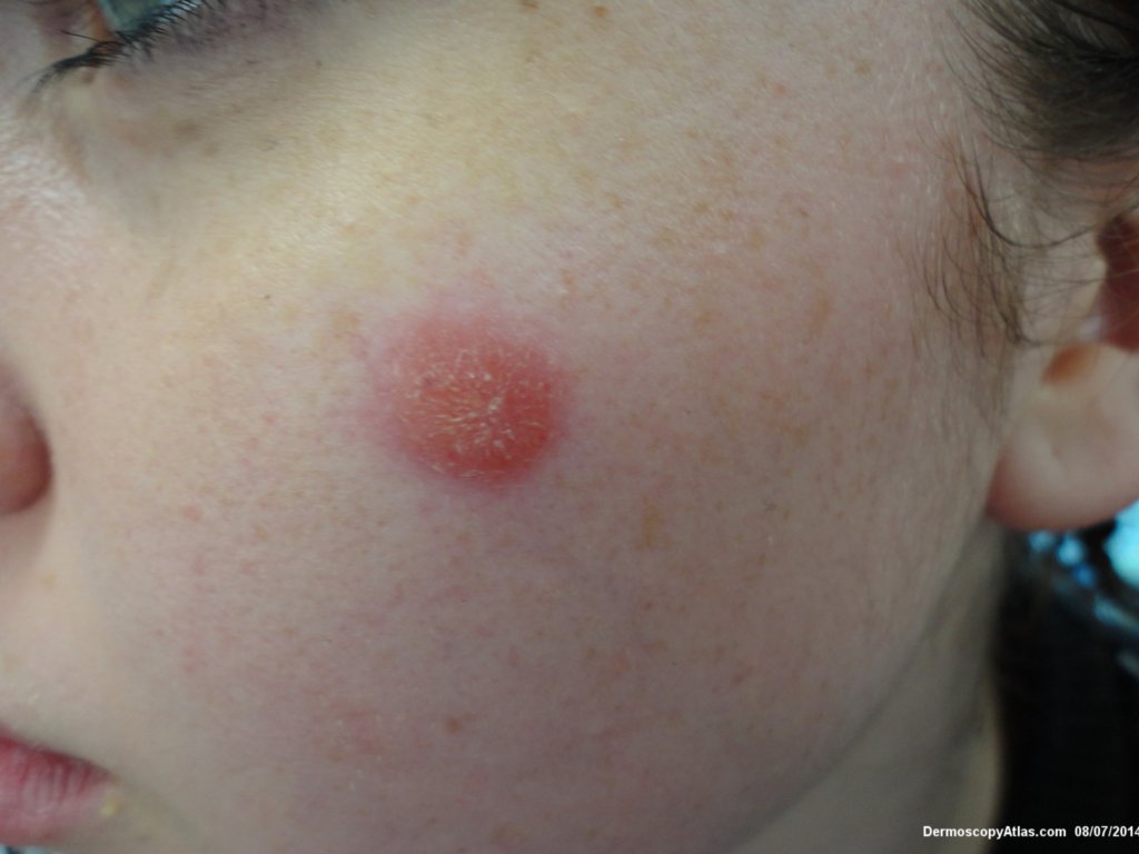
Site: Cheek
Diagnosis: Discoid lupus erythematosus
Sex: F
Age: 23
Type: Dermlite Polarised
Submitted By: Ian McColl
Description: Note the white keratin clods and dots
History:
This young girl had a solitary plaque on her cheek for 3 months. It did not resolve with cryotherapy. A biopsy was reported as showing a lichenoid reaction but this was discoid lupus erythematosus. The white clods and dots are keratin in the follicular orifices, part of follicular plugging seen in discoid lupus. It responded quickly to diluted Kenacort intralesionally plus sun protection and intermittent stronger topical steroid cream carefully applied.