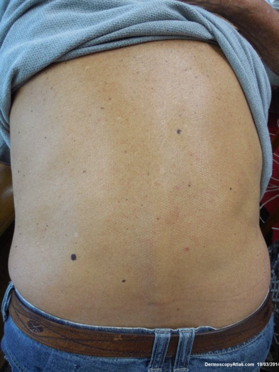
Loading
TeleDermConsult-logo
  • Recent Submissions
  • Search
  • Diagnosis
  • Quiz
  • Image comparison
  • Submit
  • Teaching Cases
  • Index
  • About

<< Back to Diagnosis Page

Image Number #3346 (Dysplastic Junctional Lentiginous Nevus)

Site: Back

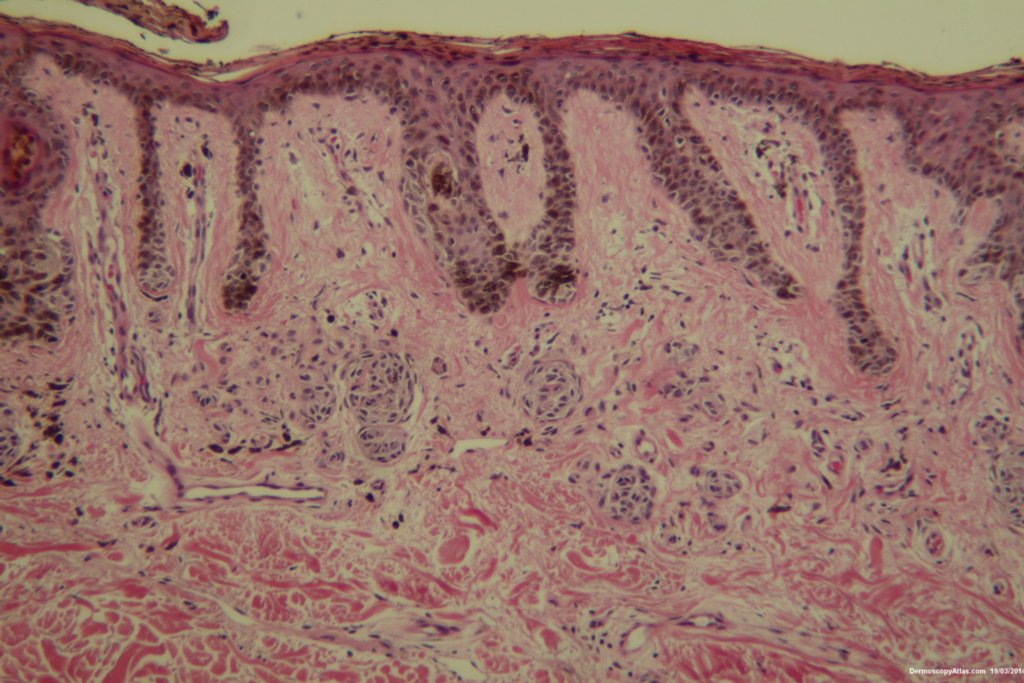
Diagnosis: Dysplastic Junctional Lentiginous Nevus

Sex: M

Age: 73

Type: Heine

Submitted By: Ian McColl

View Full Size Image

Click Here to View Details

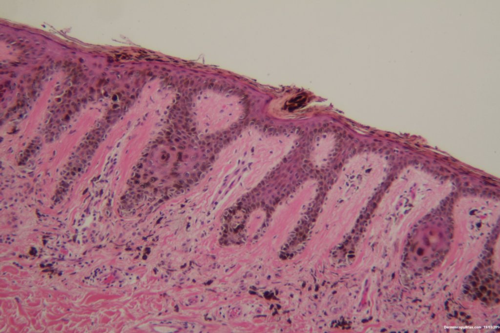
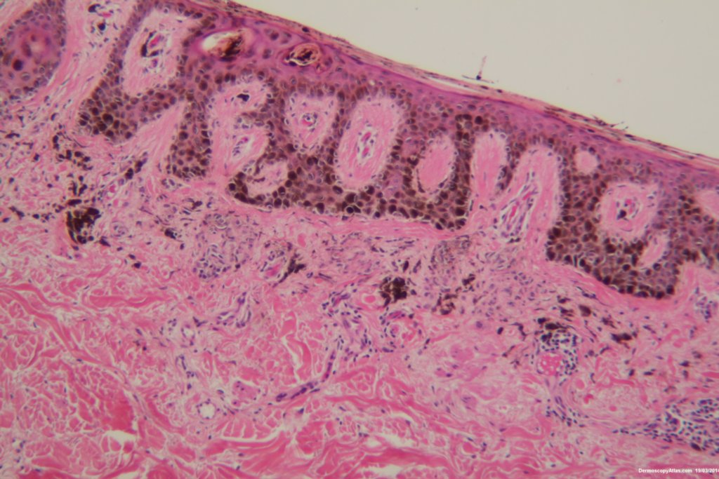
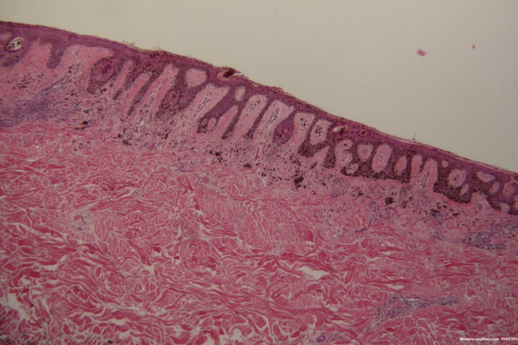
Description: More prominent normal nevus cells in the dermis. Some proliferation of melanocytes along the basal layer.

History:

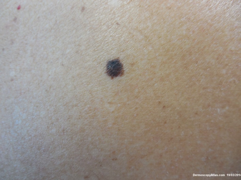
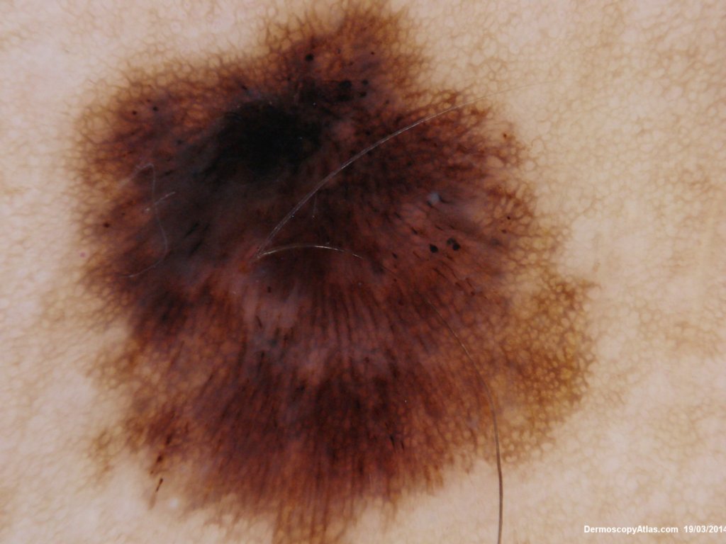
 An elderly male asked about the pigmented lesion on his lower back. The dermatoscopy showed internal lines and dark clods with a central dark structureless area. Histology showed a dysplastic junctional lentiginous nevus.

View a video of this case


DermNetNZ   eMedicine   PubMed   Dermatology Online   Archives   JAAD for "Dysplastic Junctional Lentiginous Nevus"

  • Recent Submissions
  • Search
  • Diagnosis
  • Quiz
  • Image comparison
  • Submit an Image
  • Teaching Cases
  • Index
  • About

Copyright 2022. Dermoscopy Atlas. ALL Rights Reserved.

Change Image