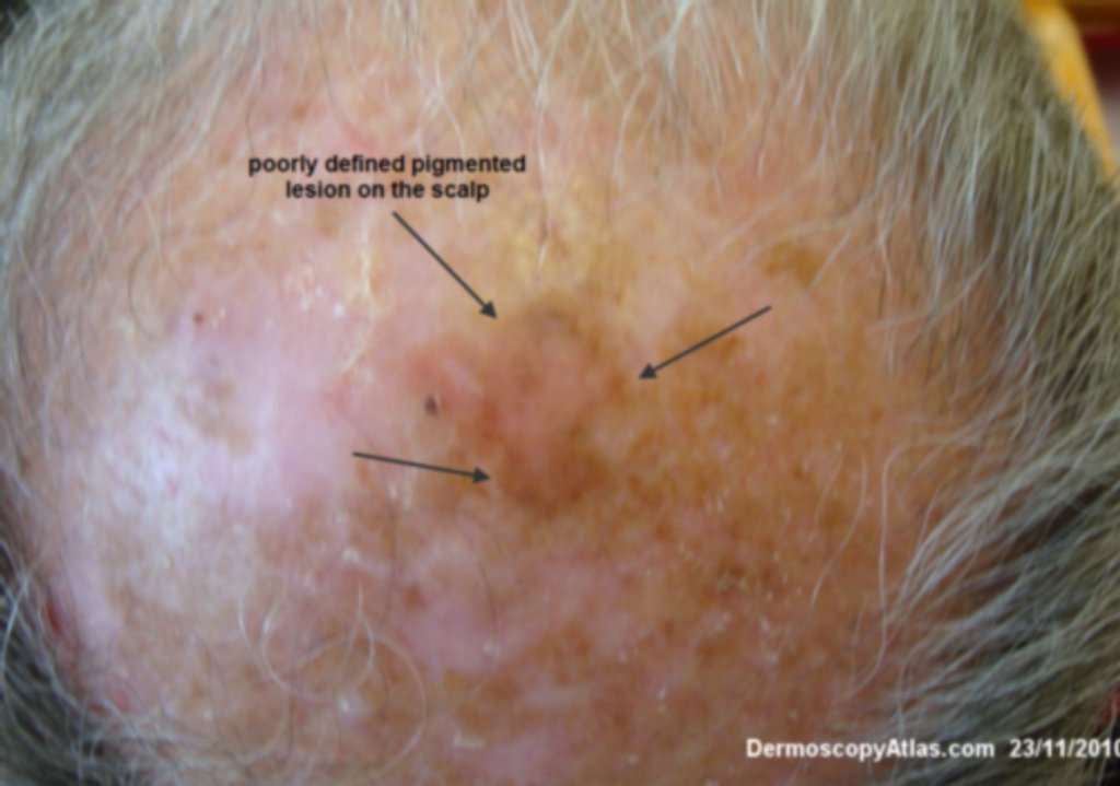
Loading
TeleDermConsult-logo
  • Recent Submissions
  • Search
  • Diagnosis
  • Quiz
  • Image comparison
  • Submit
  • Teaching Cases
  • Index
  • About

<< Back to Diagnosis Page

Image Number #3182 (Pigmented Intraepidermal carcinoma)

Site: Scalp

Diagnosis: Pigmented Intraepidermal carcinoma

Sex: M

Age: 70

Type: Dermlite Polarised

Submitted By: Ian McColl

View Full Size Image

Click Here to View Details

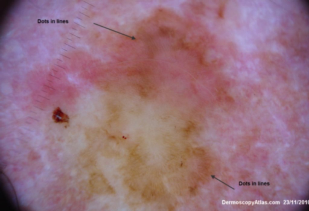
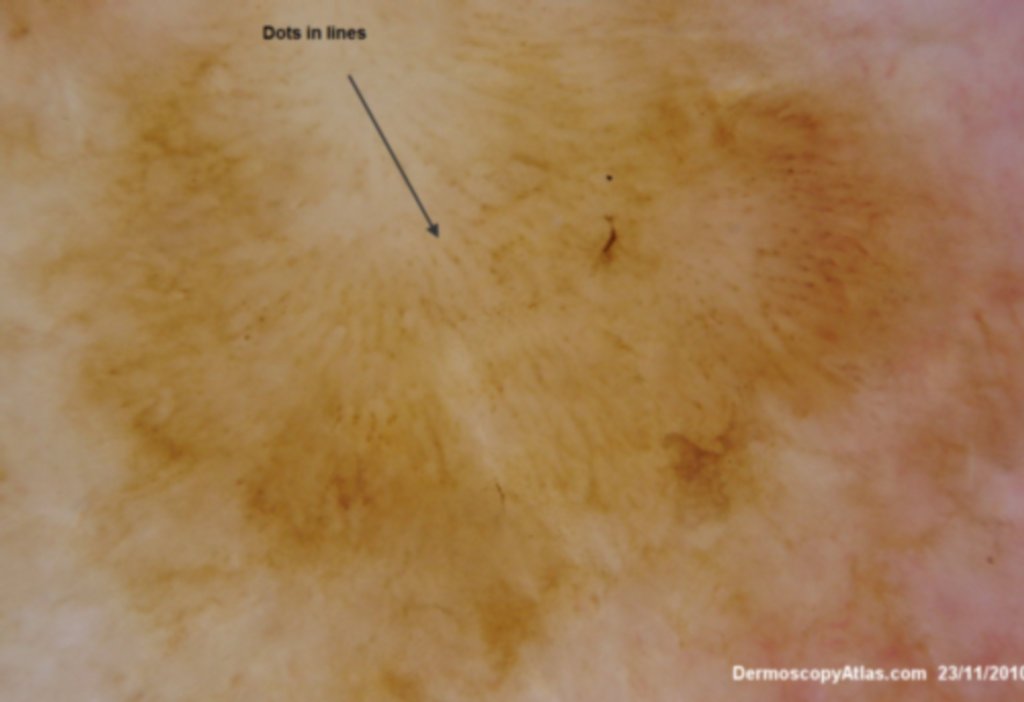
Description: Dots in lines These are more obvious in this magnified view.

History:

This poorly defined pigmented lesion on the scalp was noted on a skin examination. The area was shave biopsied and shown to be a pigmented intraepidermal carcinoma. It shows dots in lines which is a classic feature of this condition and in one image also shows the coiled vessels which are again typical of SCC in situ. He was treated with efudix cream.

DermNetNZ   eMedicine   PubMed   Dermatology Online   Archives   JAAD for "Pigmented Intraepidermal carcinoma"

  • Recent Submissions
  • Search
  • Diagnosis
  • Quiz
  • Image comparison
  • Submit an Image
  • Teaching Cases
  • Index
  • About

Copyright 2022. Dermoscopy Atlas. ALL Rights Reserved.

Change Image