
Site: Back
Diagnosis: Lentigo Maligna
Sex: M
Age: 80
Type: Heine
Submitted By: Jeffrey Keir
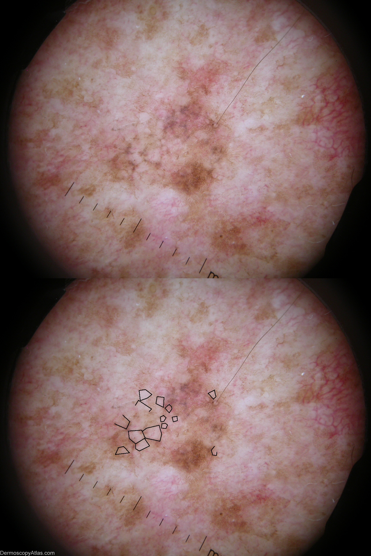
Description: A 7.5 mm diameter lentiginous melanoma in situ on the upper back of an 80 y.o. male. Note the background pattern of light brown network with some “fingerprinting”, a pattern described as present in solar lentigo; but the sharply demarcated scalloped border of solar lentigo is absent. To the left of the image there are obvious large polygons defined by grey lines (formed of aggregations of grey dots). More subtle polygons defined by darker surrounding pigment are present elsewhere
History:
Large Polygons - a dermoscopic feature of Lentiginous Melanoma
see more detailed text in PDF here
21/27 non-facial lentiginous melanomas detected over a 2 year period had these structures.
These polygonal shapes may be obvious and complete or subtle and incomplete. They are defined by darker grey or brown relatively straight lines, or by darker (sometimes only barely so) surrounding lesional pigment or a combination of the two. They are much larger than the rhomboidal structures seen in facial lentigo maligna and may be rhomboidal, pentagonal, or hexagonal. In some cases the large polygons do appear to be centred on follicular openings, as in lentigo maligna of the face, but this is not a constant.
I have termed these shapes “large polygons”, in contrast to the smaller rhomboidal structures seen in facial lentigo maligna.
Large polygons usually occurred within a pigmented lesion that had lentigo-like pigment patterns, but lacking the sharply defined scalloped edge usually seen with solar lentigo.