
Site: Other
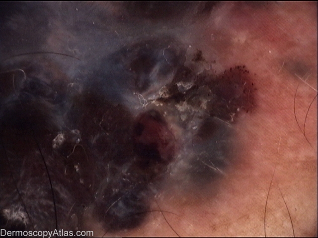
Diagnosis: Malignant blue nevus
Sex: M
Age: 56
Type: Molemax
Submitted By:
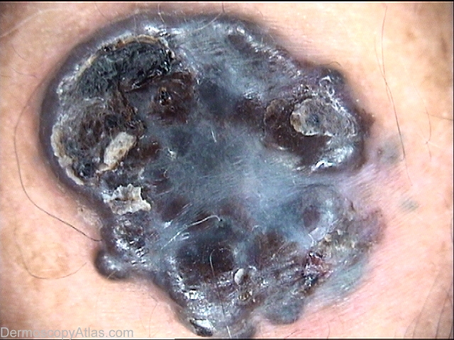
Description: Blue-gray nodular lesion with bluish to white-gray structureless pigmentation.
History: A 56 -year-old man came because he noticed the presence of a "nevus" for a short period of time. Clinically and dermoscopically it was suspicious of melanoma and a surgical excision was performed. Histological examination confirmed a diagnosis of MBN with polygonal melanocytes with prominent nucleoli and a few mitotic figures, predominatly in the reticular dermis. Breslow thickness was 11mm, Clark V with diffuse expression of an immunohistochemical stain for CD34.