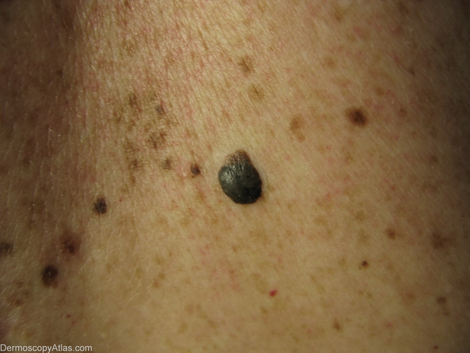
Loading
TeleDermConsult-logo
  • Recent Submissions
  • Search
  • Diagnosis
  • Quiz
  • Image comparison
  • Submit
  • Teaching Cases
  • Index
  • About

<< Back to Diagnosis Page

Image Number #3136 (Seborrhoeic keratosis)

Site: Back

Diagnosis: Seborrhoeic keratosis

Sex: F

Age: 47

Type: Heine

Submitted By: Ian McColl

View Full Size Image

Click Here to View Details

Description: Dark raised pigmented lesion on the back

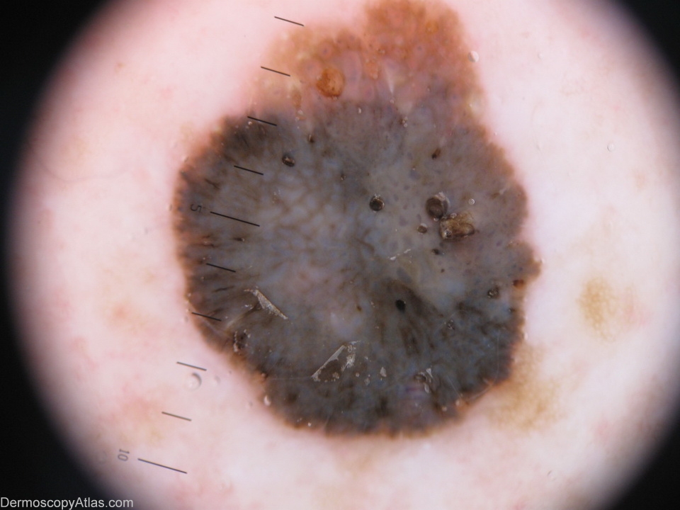
History: The thicker dark seborrhoeic keratosis will often have a bluish tinge to it under the dermatoscope presumably because of the depth of melanin pigment in a thick lesion. All this pigment is in epidermal cells whereas we usually associate the colour blue with melanin pigment or nests deep in the dermis of melanocytic lesions.

DermNetNZ   eMedicine   PubMed   Dermatology Online   Archives   JAAD for "Seborrhoeic keratosis"

  • Recent Submissions
  • Search
  • Diagnosis
  • Quiz
  • Image comparison
  • Submit an Image
  • Teaching Cases
  • Index
  • About

Copyright 2022. Dermoscopy Atlas. ALL Rights Reserved.

Change Image