
Site: Breast
Diagnosis: Nevus dermal
Sex: F
Age: 27
Type: Heine
Submitted By: Greg Canning
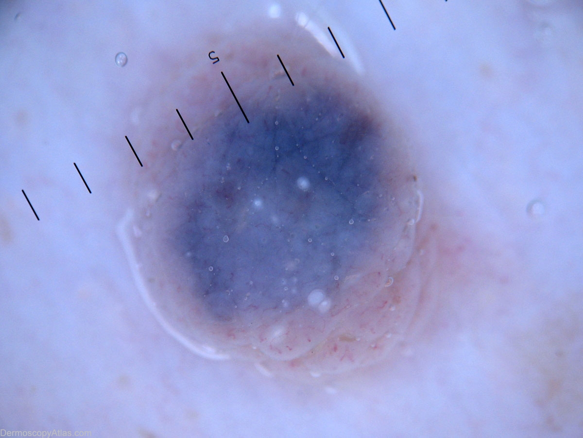
Description: Dermoscopy : Central blue homogenous area, peripheral pink structureless area with linear and comma like vessels, and a few sattered milia like cysts.
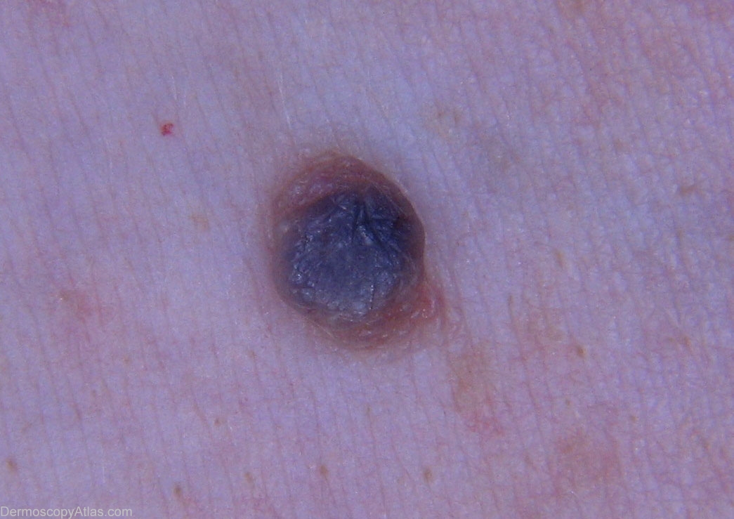
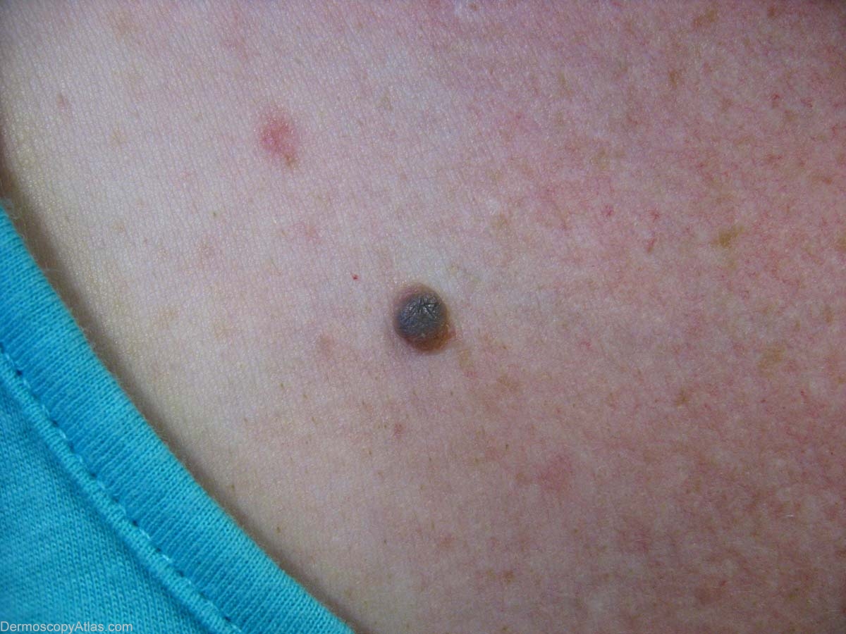
History: This 27 yr old woman presented with an enlarging lesion with cetral darkening of colour. Clinically it was felt to be a combined nevus. Histpathology was reported : Sections show a lesion which is essentially a polypoid INTRADERMAL NAEVUS. However, the morphology differs in different parts of the lesion. Superficially, laterally and in the deep aspect of the lesion the appearances are those of ordinary intradermal naevus. Centrally however there is substantial sclerosis, naeval cells are larger with voluminous cytoplasm and finely divided melanin pigment, there is sclerosis and numerous melanophages which are heavily pigmented are present in this area. The heavily pigmented melanophages among the nests of naeval cells are somewhat reminiscent of a deep penetrating naevus, but the cytology of the naeval cells themselves are not typical for that diagnosis. There is no evidence of malignancy. The excision appears complete.

