
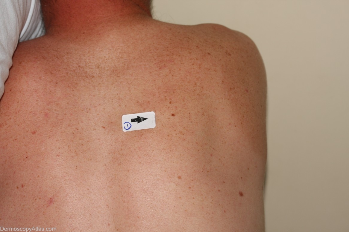
Site: Back
Diagnosis: Melanoma in situ
Sex: M
Age: 28
Type: Dermlite Non Polarised
Submitted By: Cliff Rosendahl
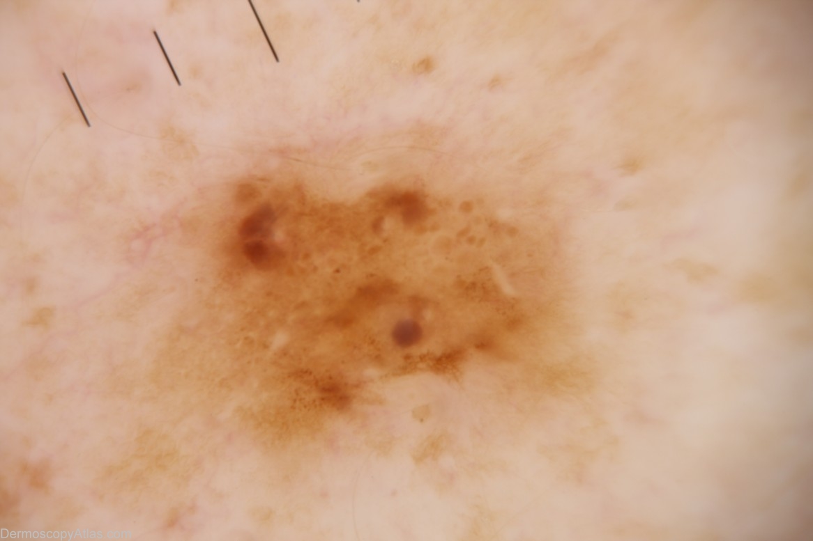
Description: Dermoscopy image - Pattern - Clods and dots. Colours - Brown and blue-grey. Clues. No strong clues. The clods are varied in size and randomly distributed. Brown dots are focal. There isc a hint of inverse network
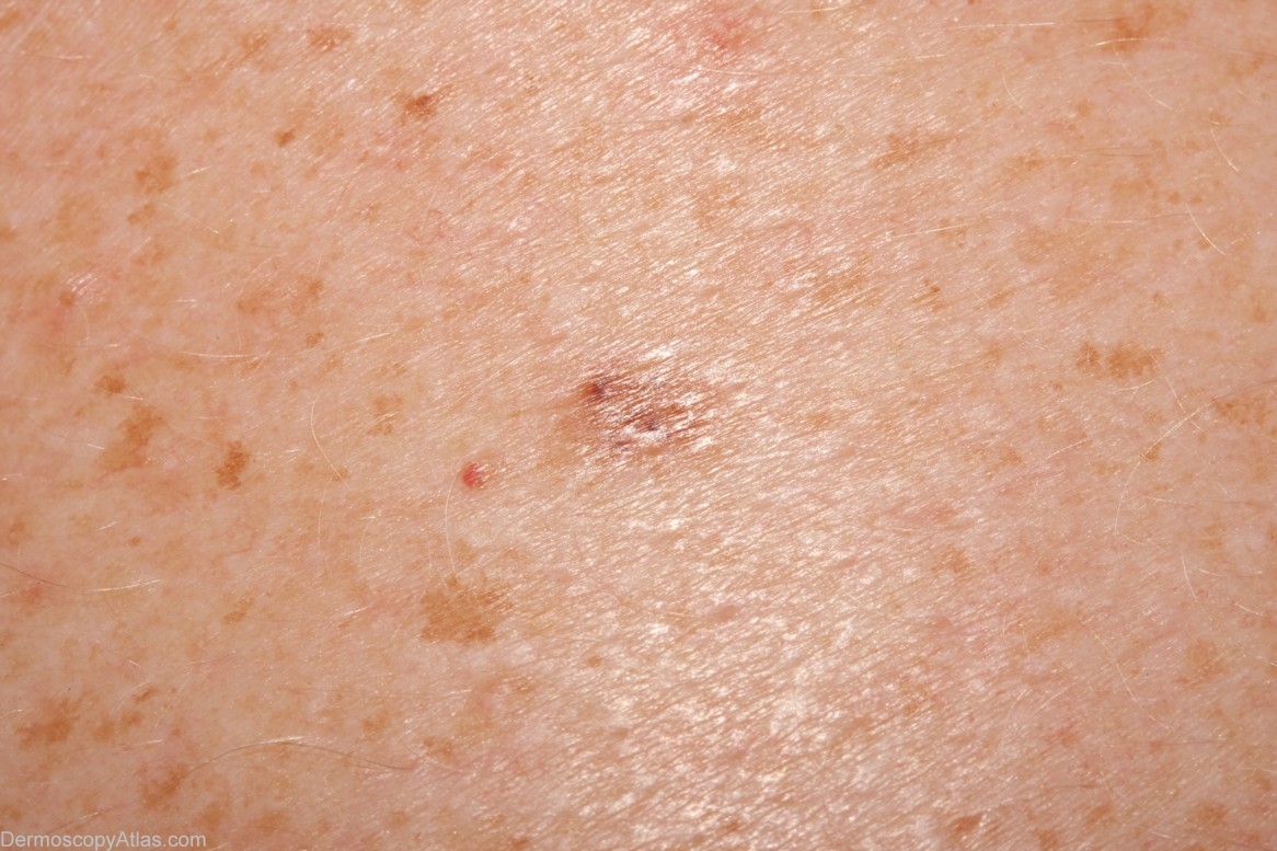
History: This 28 year old concreter presented for his first ever skin examination. This lesion was noticed and imaged with the intention of monitoring. On viewing the image the author decided to excise it mainly because of a suggestion of inverse network and focal brown dots.