
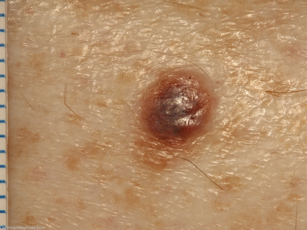
Site: Shins
Diagnosis: Melanoma invasive
Sex: F
Age: 49
Type: Dermlite Non Polarised
Submitted By: Alan Cameron
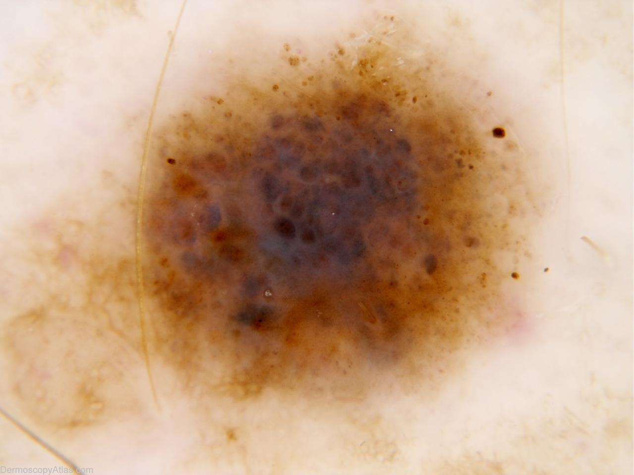
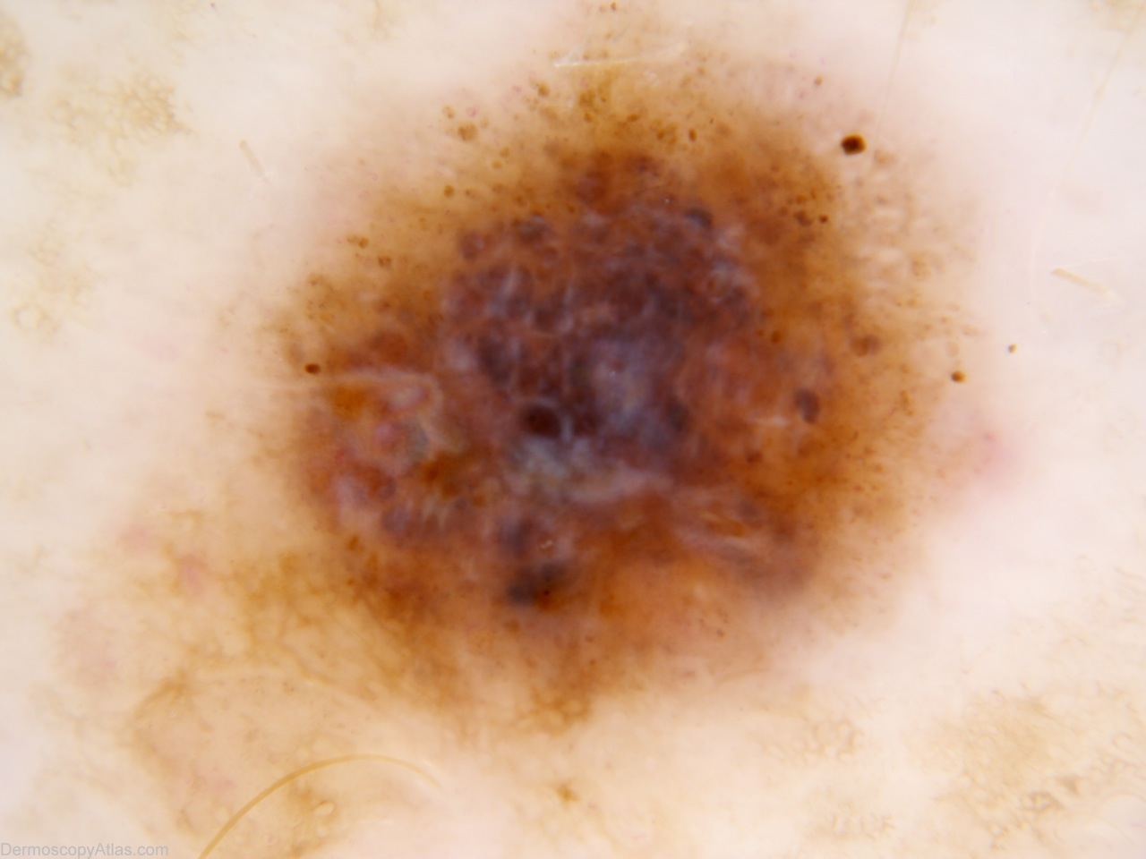
Description: Clinical macro is quite innocuous. mm scale on left.
History: Enlarging pigmented lesion over hte last 12 months. No past history, but mother died of melanoma. Histology reported as: The shave shows level 2 superficial spreading malignant melanoma. It is non-ulcerated and has a Breslow thickness of 0.5mm. There is an occasional dermal mitosis present indicating that the lesion is in vertical growth plane. No lymphovascular or perineural infiltration is identified. There is evidence of focal regression.