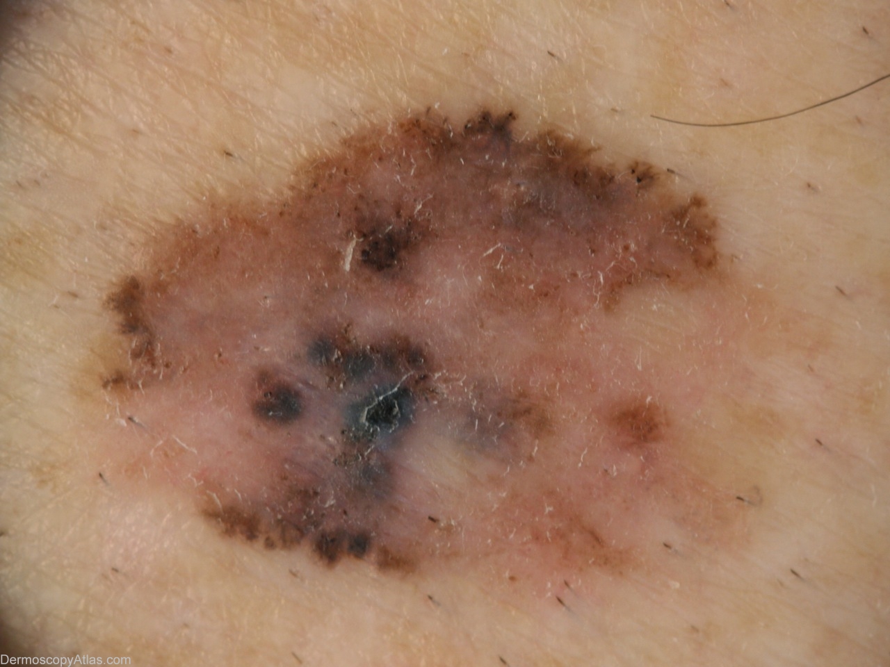
Loading
TeleDermConsult-logo
  • Recent Submissions
  • Search
  • Diagnosis
  • Quiz
  • Image comparison
  • Submit
  • Teaching Cases
  • Index
  • About

<< Back to Diagnosis Page

Image Number #3032 (Melanoma invasive)

Site: Thigh

Diagnosis: Melanoma invasive

Sex: M

Age: 64

Type: Dermlite Non Polarised

Submitted By: Alan Cameron

View Full Size Image

Click Here to View Details

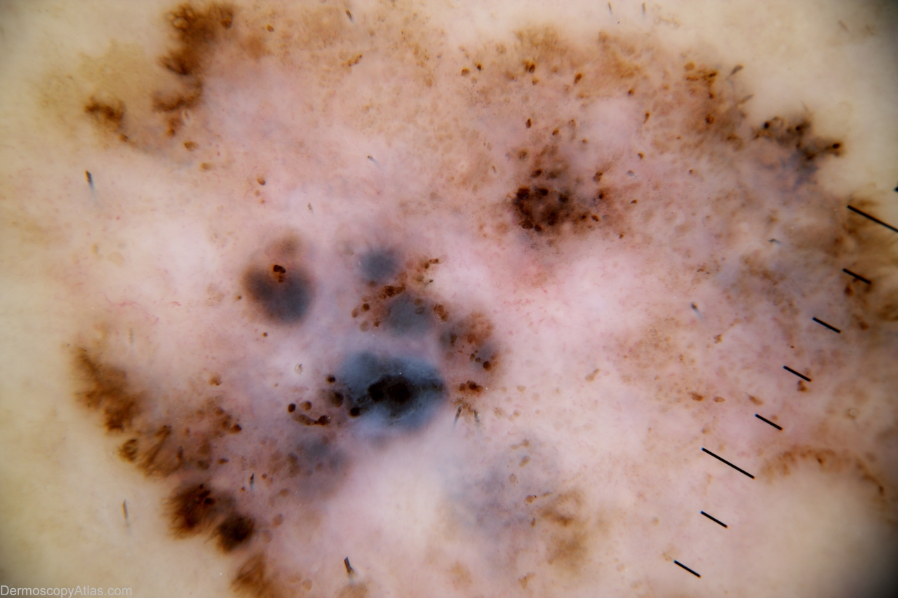
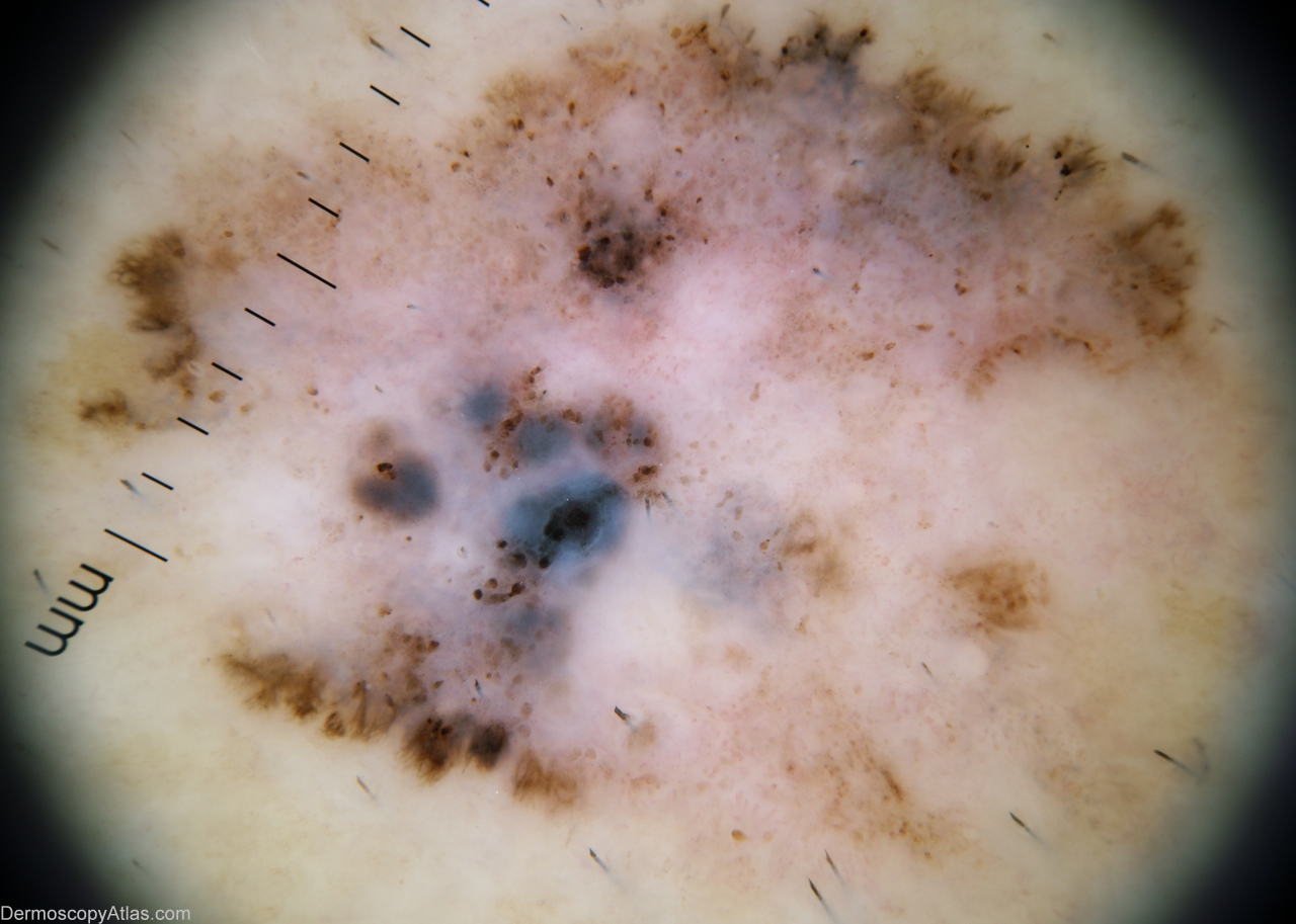
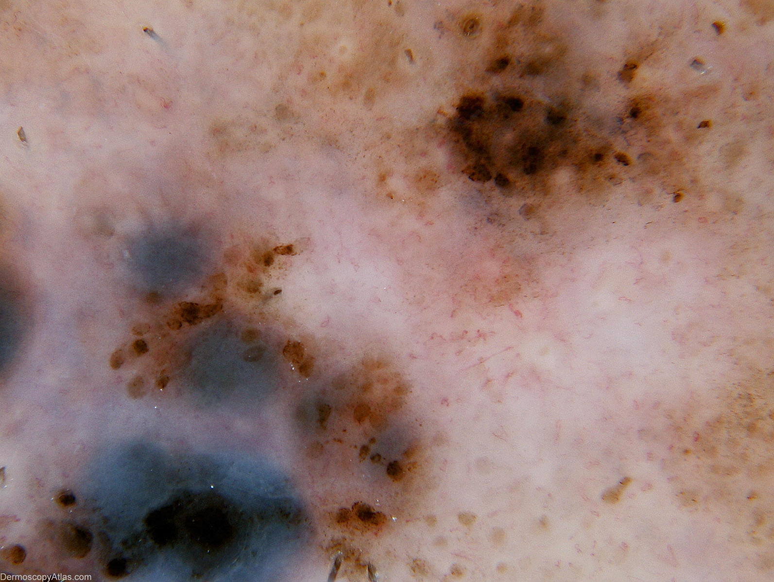
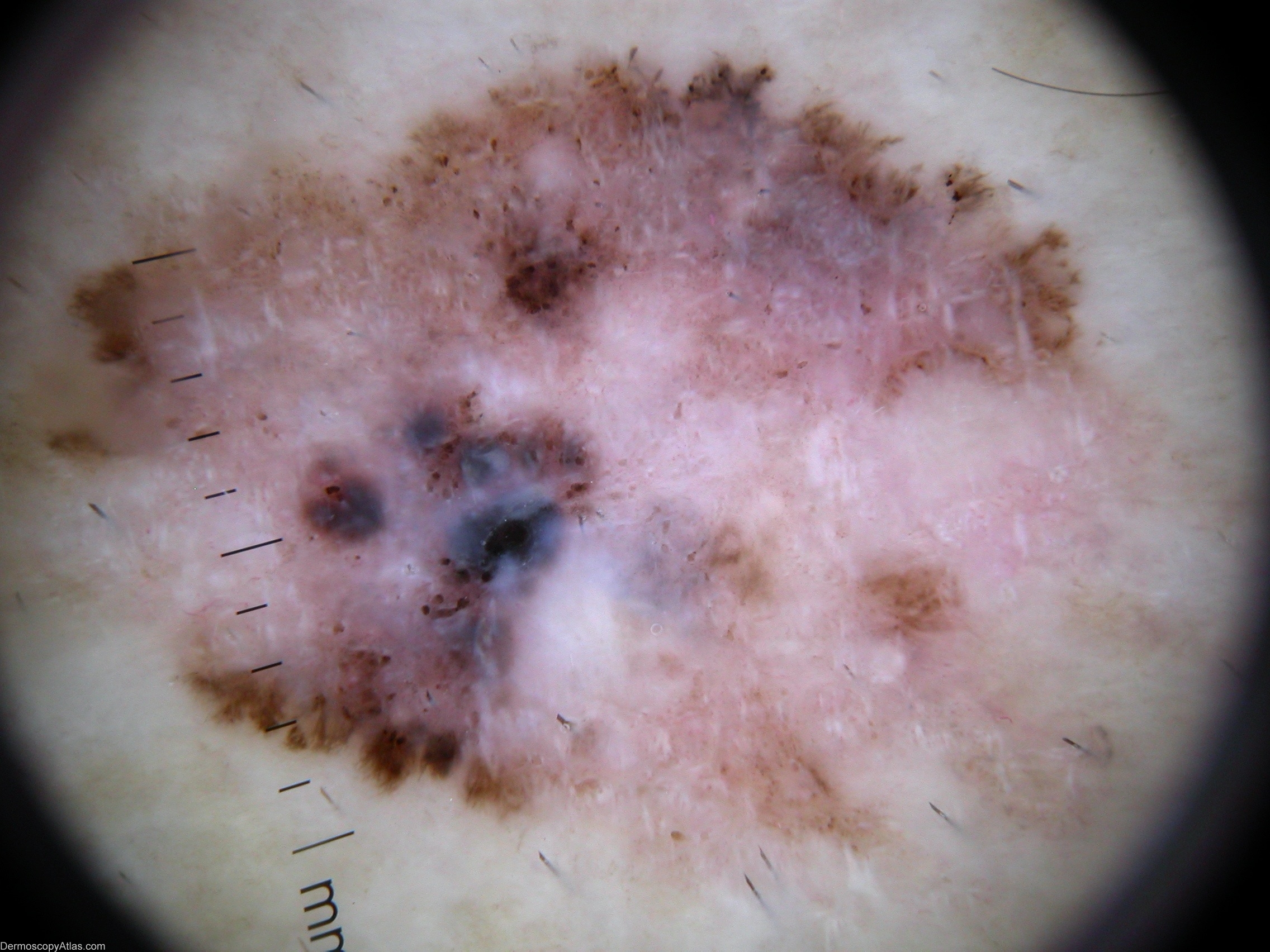
Description: Macro shows well defined variegated plaque

History: Progressively enlarging pigmented lesion on thigh. Past history BCCs. Histolgy Reported as: Sections confirm level 3 (vertical growth phase) superficial spreading malignant melanoma. It is non-ulcerated and has a Breslow thickness of approximately 0.7mm. Occasional dermal mitoses are evident. There is evidence of both active and established past regression. No lymphovascular or perineural infiltration is seen. There is an underlying benign naevus present.

DermNetNZ   eMedicine   PubMed   Dermatology Online   Archives   JAAD for "Melanoma invasive"

  • Recent Submissions
  • Search
  • Diagnosis
  • Quiz
  • Image comparison
  • Submit an Image
  • Teaching Cases
  • Index
  • About

Copyright 2022. Dermoscopy Atlas. ALL Rights Reserved.

Change Image