
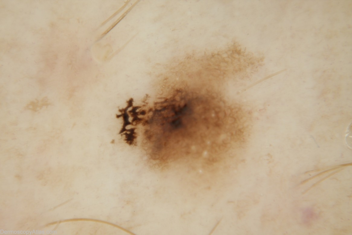
Site: Leg
Diagnosis: Dysplastic nevus
Sex: M
Age: 44
Type: Dermlite Non Polarised
Submitted By: Cliff Rosendahl
Description: dermoscopic image - This lesion contains both lines reticular (brown circles) and lines curved which meant a confident melanocytic diagnosis was not forthcoming. White clods (milia cysts) further suggested seborrhoeic keratosis. The eccentric pigment was alarmingly unusual.
History: The very small lesion was observed at a routine skin check. The eccentric focus of pigment attracted attention to it. The author thought that if it was not melanocytic it would be a seborrhoeic keratosis (provisional diagnosis) but if melanocytic then melanoma needed to be excluded. Histology revealed it to be a (moderately dysplastic) naevus.