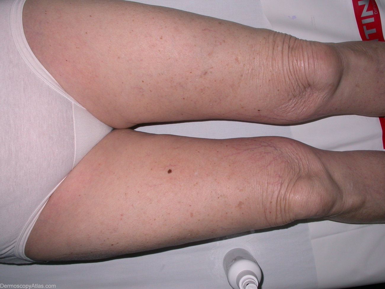
Loading
TeleDermConsult-logo
  • Recent Submissions
  • Search
  • Diagnosis
  • Quiz
  • Image comparison
  • Submit
  • Teaching Cases
  • Index
  • About

<< Back to Diagnosis Page

Image Number #2957 (Melanoma in situ)

Site: Thigh

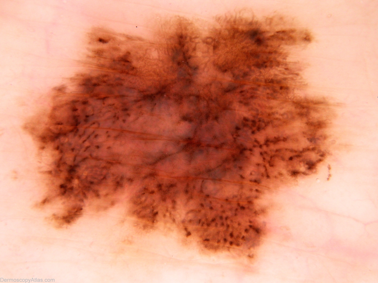
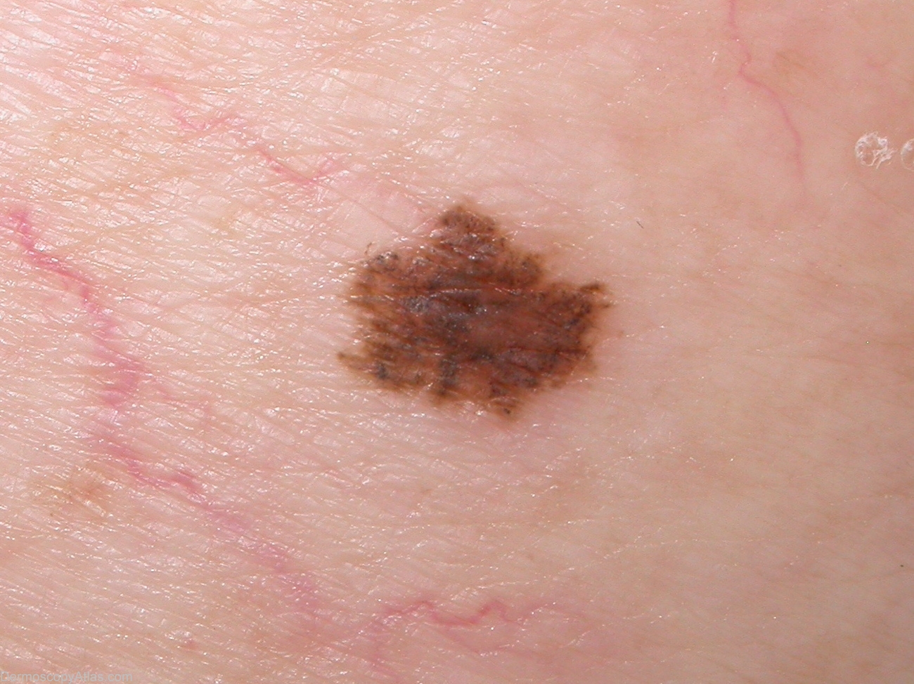
Diagnosis: Melanoma in situ

Sex: F

Age: 79

Type: Dermlite Non Polarised

Submitted By: Alan Cameron

View Full Size Image

Click Here to View Details

Description: Wide view shows solitary pigmented lesion.

History: 79 year old woman with past history of BCCs. This lesion noted at routine review. Histology reported as; Sections show level 1 superficial spreading malignant melanoma with regression arising a junctional and lentiginous dysplastic naevus. There is no ulceration and no definite dermal invasion is seen.

DermNetNZ   eMedicine   PubMed   Dermatology Online   Archives   JAAD for "Melanoma in situ"

  • Recent Submissions
  • Search
  • Diagnosis
  • Quiz
  • Image comparison
  • Submit an Image
  • Teaching Cases
  • Index
  • About

Copyright 2022. Dermoscopy Atlas. ALL Rights Reserved.

Change Image