
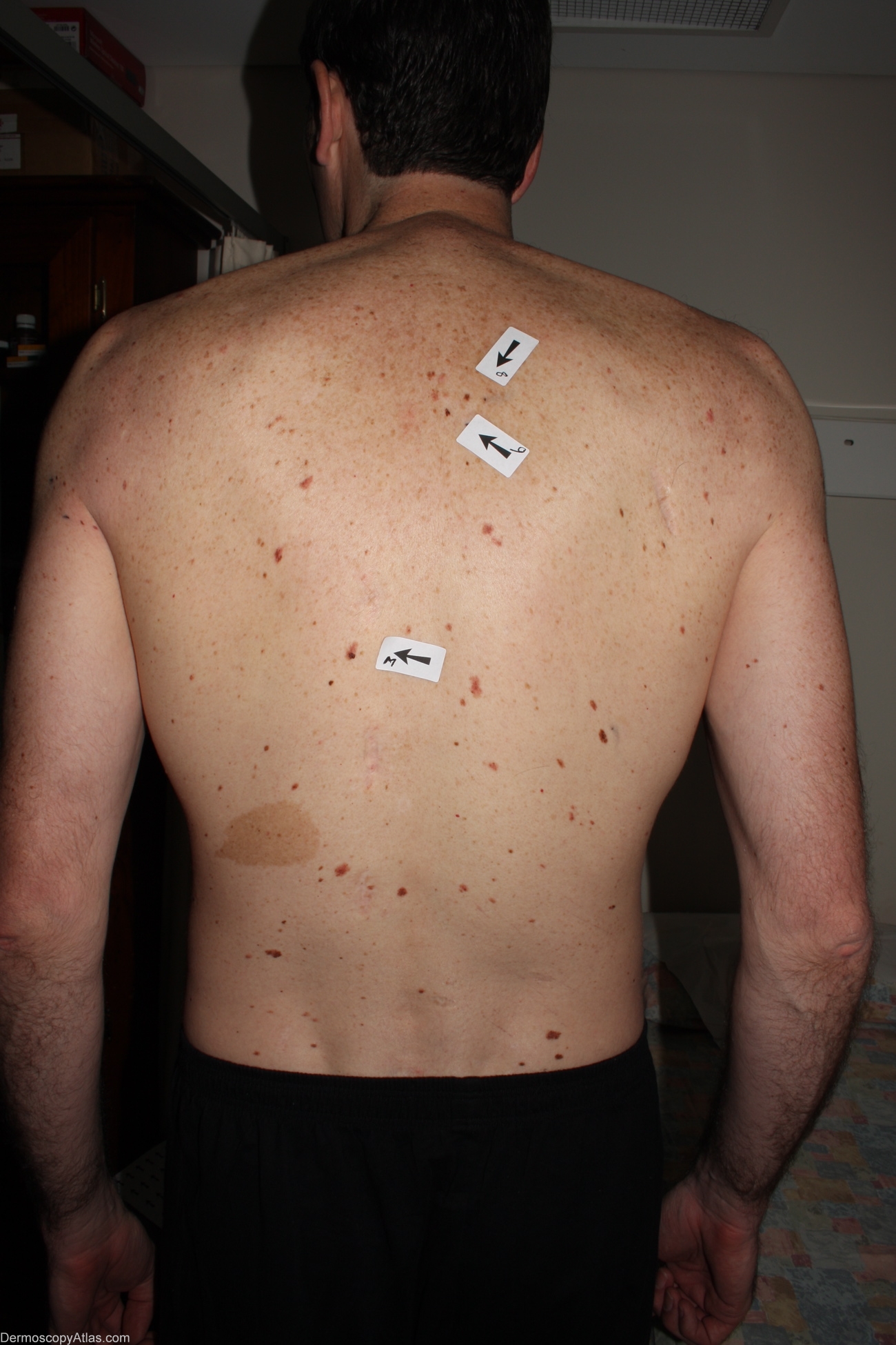
Site: Back
Diagnosis: Melanoma in situ
Sex: M
Age: 35
Type: Dermlite Non Polarised
Submitted By: Alan Cameron
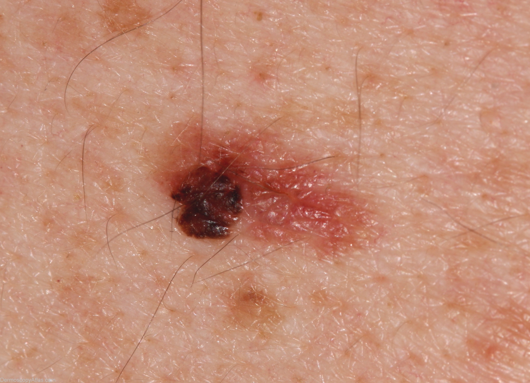
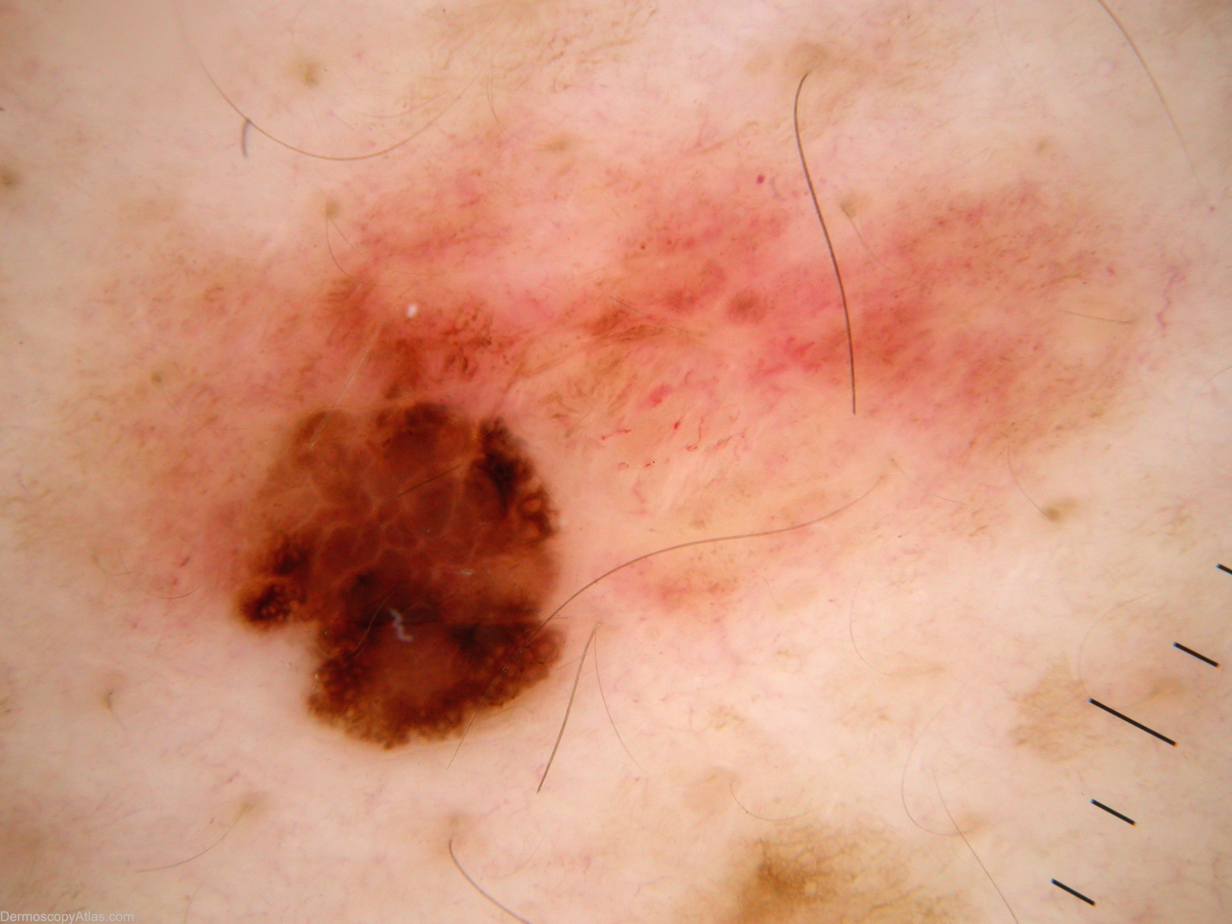
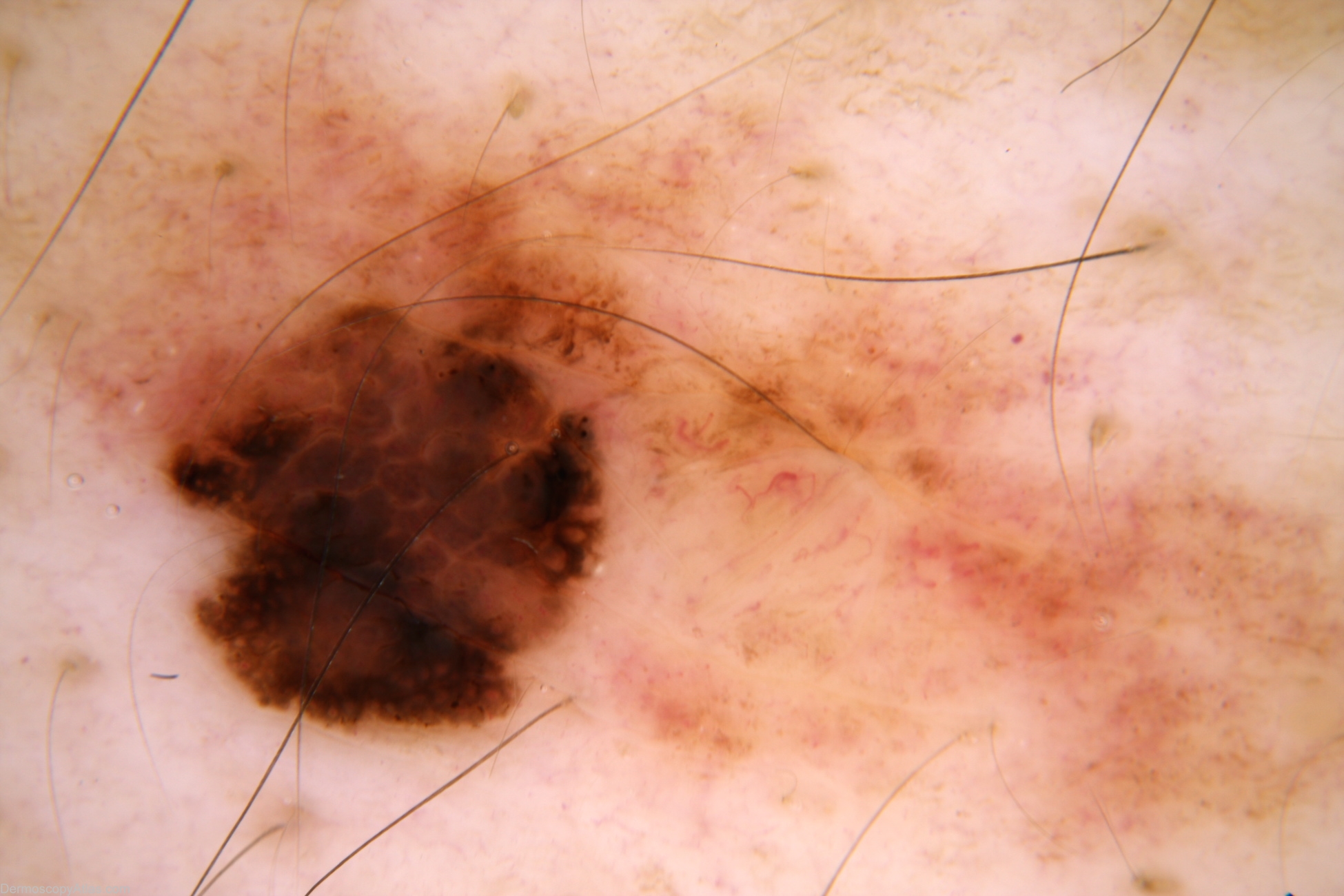
Description: Against the background of structureless tan lesion, a focus of thick lines reticular with irregular black and brown clods, and black dots peripherally (in this focus). Pattern in the superior portion of this focus is brown clods and not white lines reticular (negative network) as the lines are not lighter than surrounding skin.
History: 35 year old male with atypical mole syndrome but no past history. Referred by GP regarding this lesion. Melanoma 394 was also found at this examination. Histology reported as; Sections show a level 1 (in situ) superficial spreading melanoma arising in a dysplastic compound naevus. There is no ulceration, dermal invasion, lymphocytic infiltrate or regression.