
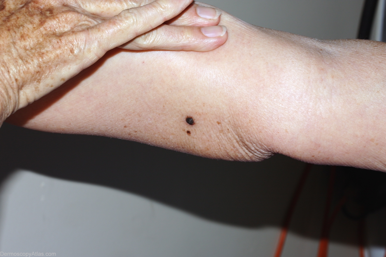
Site: Arm,upper
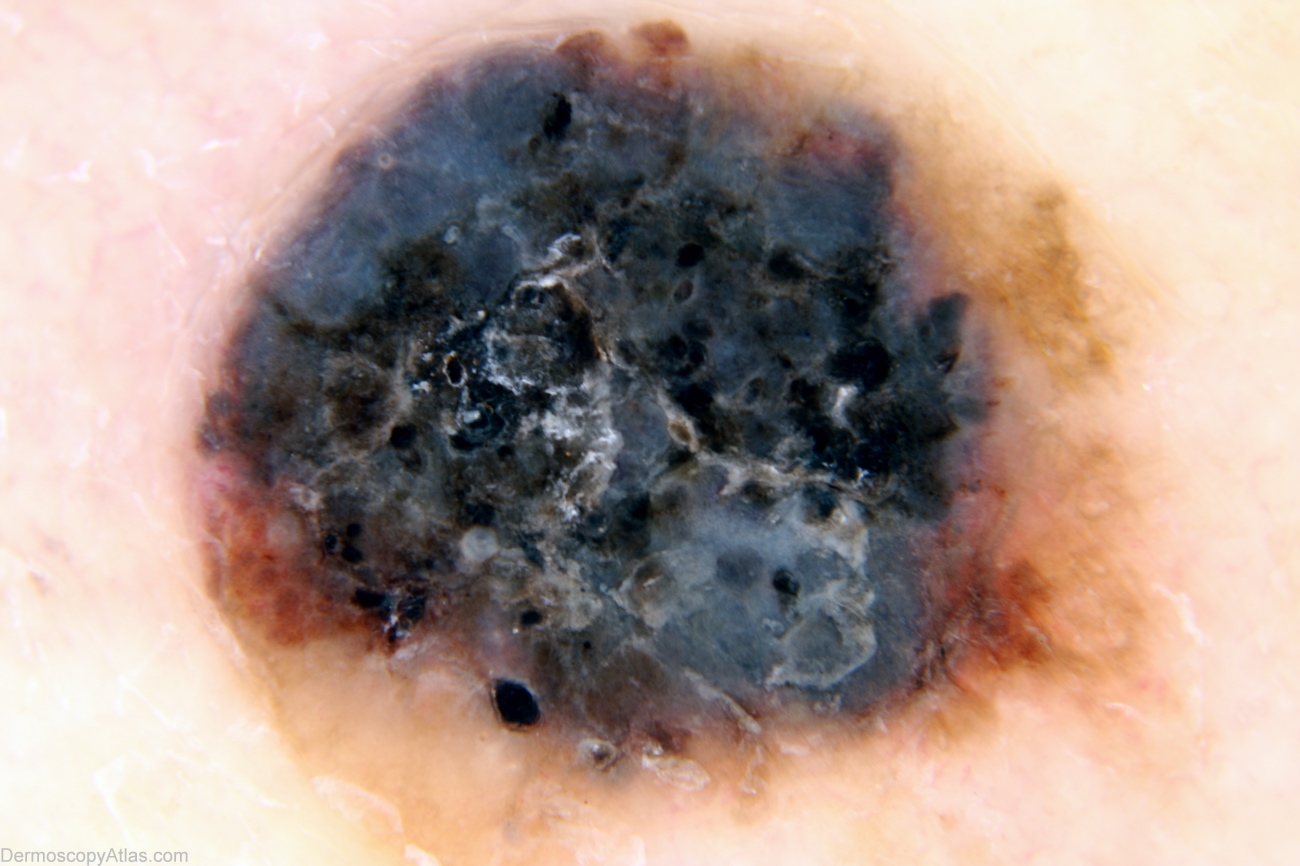
Diagnosis: Melanoma nodular
Sex: M
Age: 71
Type: Mixed
Submitted By: Alan Cameron
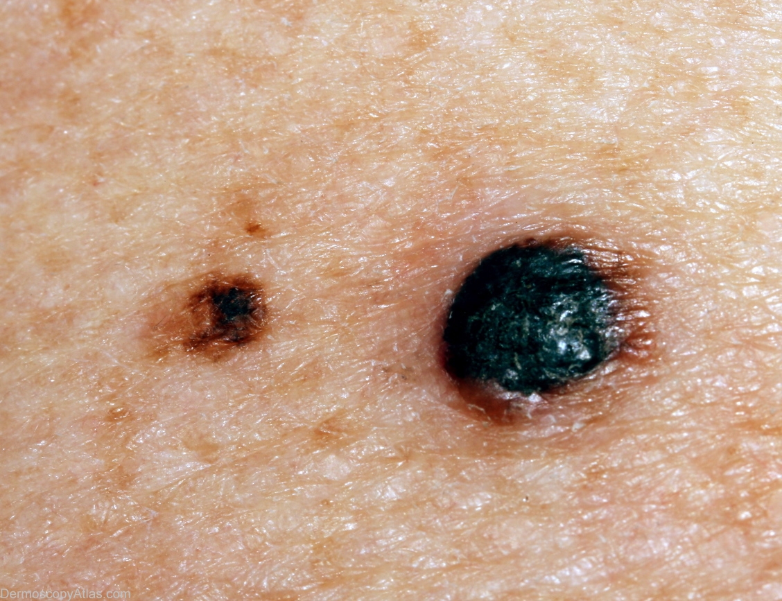
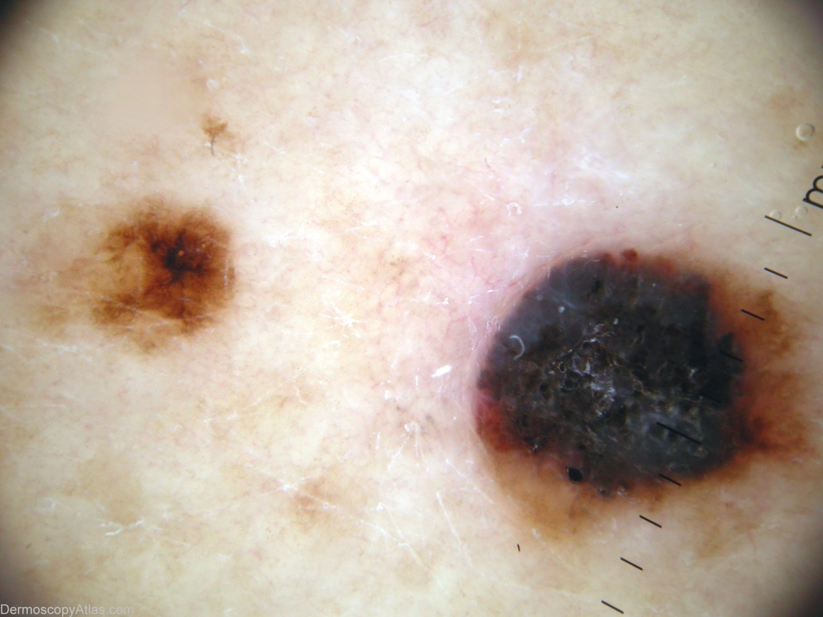
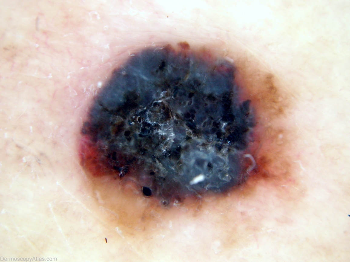
Description: Location showing small residual of previuos macule
History: History is of a macule arising on the upper arm 3 years previously, which shrank and was replaced by the nodule now apparent. There is a small part of the previous macule still visible. The histology was reported as; Sections show an early level 4 (1.5mm thick) superficial spreading melanoma. There is no ulceration. The mitotic count is low. There is a mild lymphocytic infiltrate in the base. There is no significant regression in the lesion. The other lesion is an almost completely regressed solar lentigo.