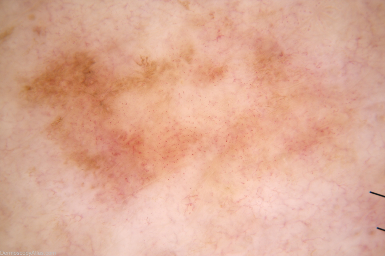
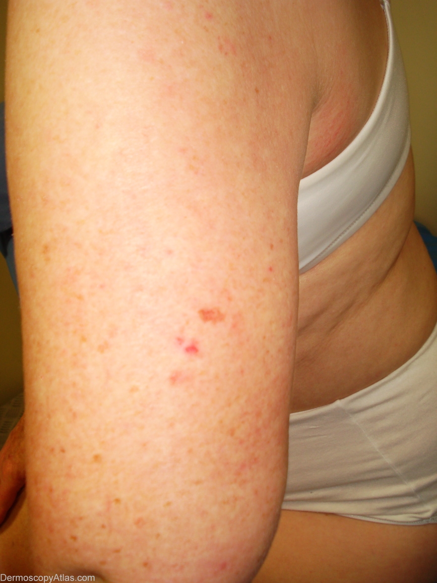
Loading
TeleDermConsult-logo
  • Recent Submissions
  • Search
  • Diagnosis
  • Quiz
  • Image comparison
  • Submit
  • Teaching Cases
  • Index
  • About

<< Back to Diagnosis Page

Image Number #2924 (Melanoma amelanotic)

Site: Arm,upper

Diagnosis: Melanoma amelanotic

Sex: F

Age: 60

Type: Mixed

Submitted By: Alan Cameron

View Full Size Image

Click Here to View Details

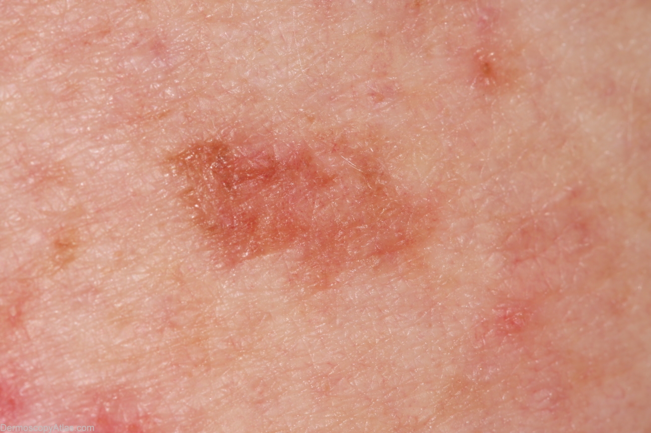
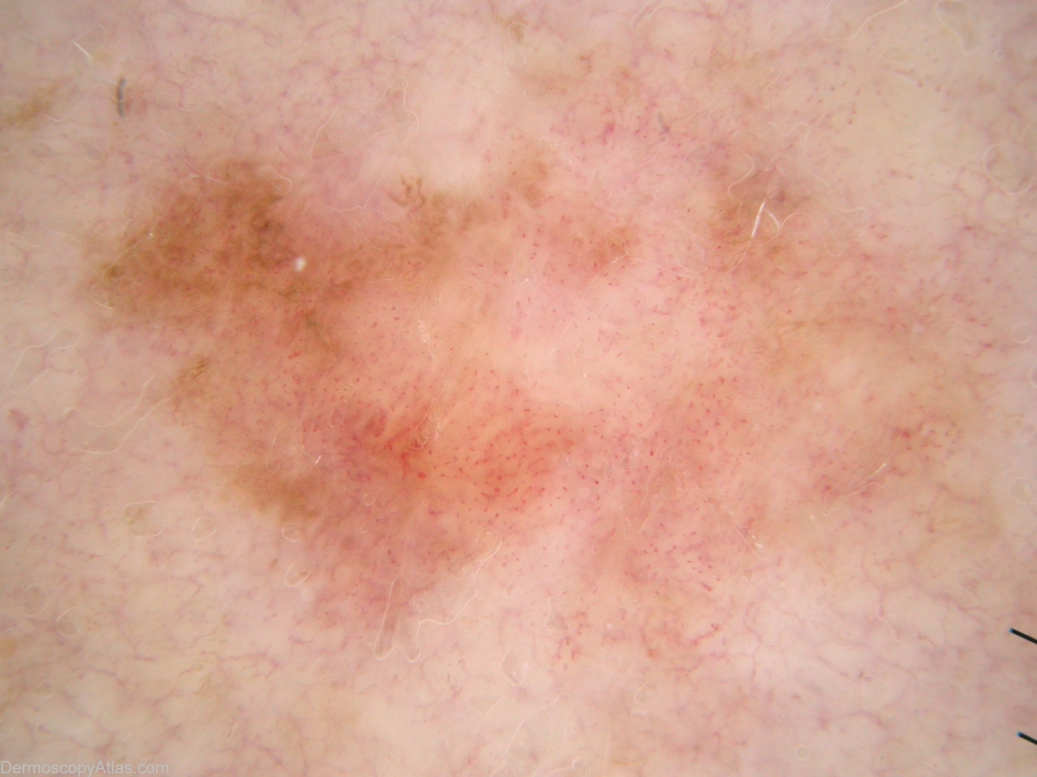
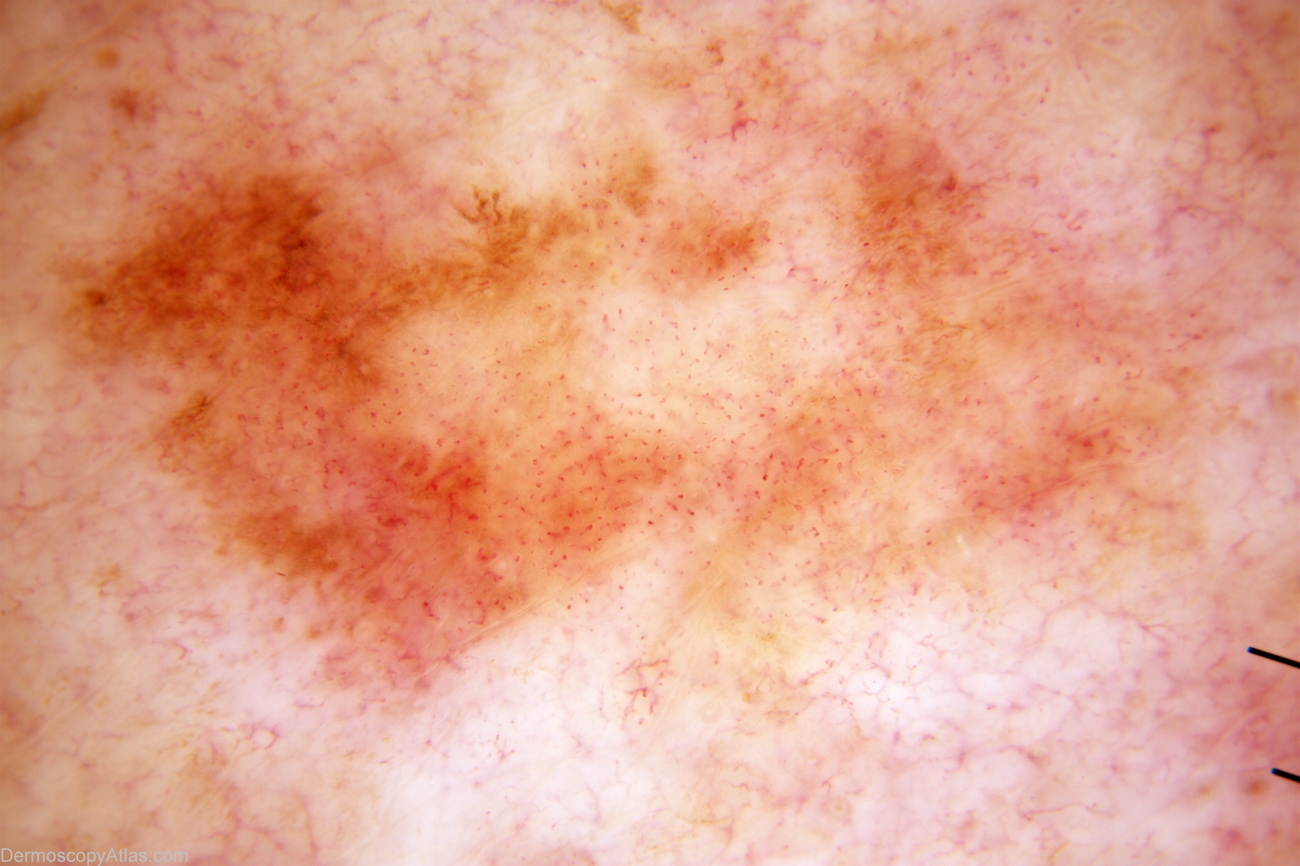
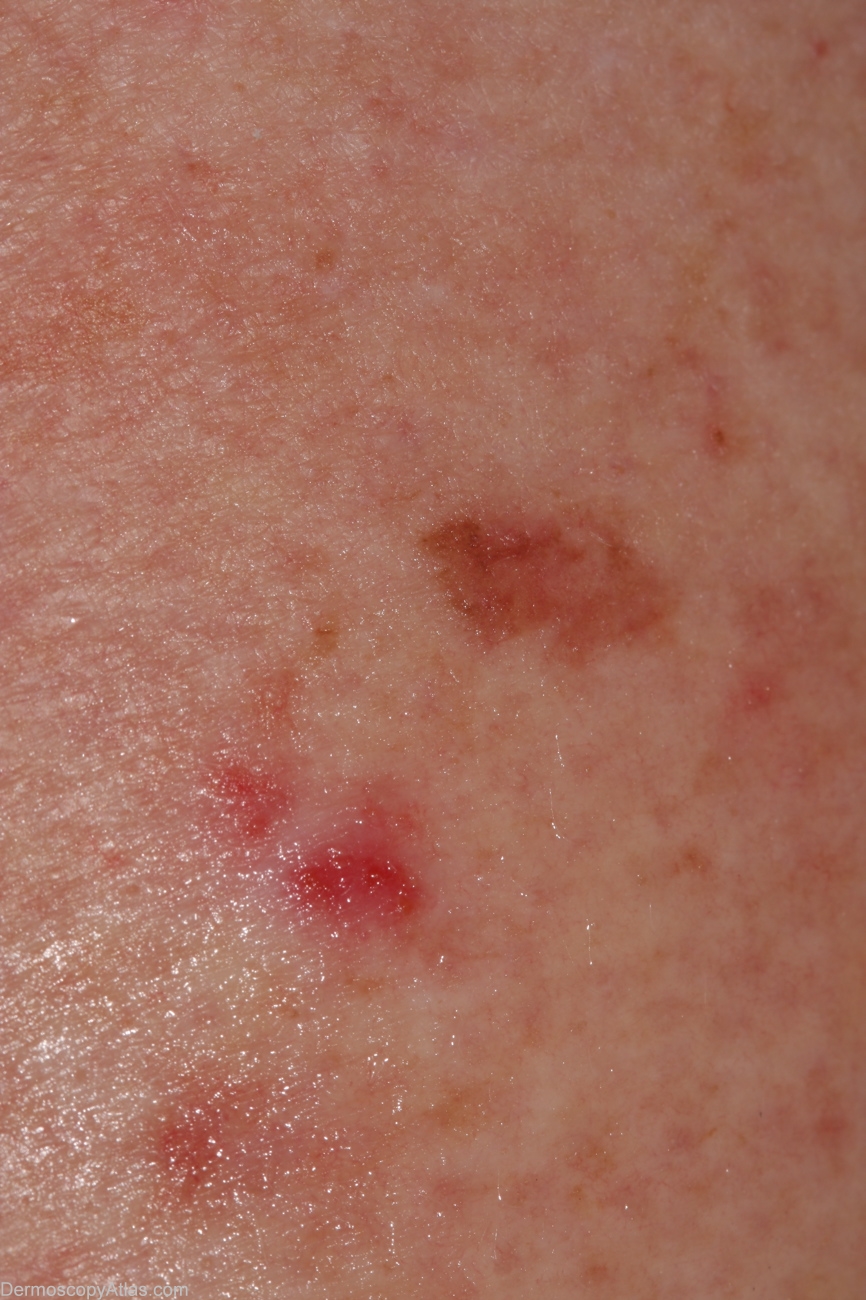
Description: Non-polarising with Dermlite Fluid. Vascular structures predominate with a few lines reticular top left (overemphased in these photos) a clue to melanocytic lesion.

History: 60 year old woman with long history of BCCs, new lesion upper arm since last check 6 months prior. Lesion today is superior of two, inferior is another BCC. Histology reported as; Sections confirm a level 1 superficial spreading malignant melanoma arising in a dysplastic naevus. There is no ulceration and no invasion is seen.

DermNetNZ   eMedicine   PubMed   Dermatology Online   Archives   JAAD for "Melanoma amelanotic"

  • Recent Submissions
  • Search
  • Diagnosis
  • Quiz
  • Image comparison
  • Submit an Image
  • Teaching Cases
  • Index
  • About

Copyright 2022. Dermoscopy Atlas. ALL Rights Reserved.

Change Image