
Site: Shins
Diagnosis: Melanoma - recurrent
Sex: F
Age: 71
Type: Dermlite Non Polarised
Submitted By: Cliff Rosendahl
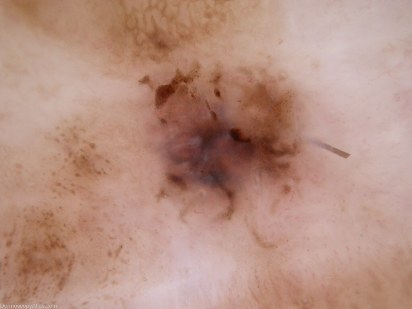
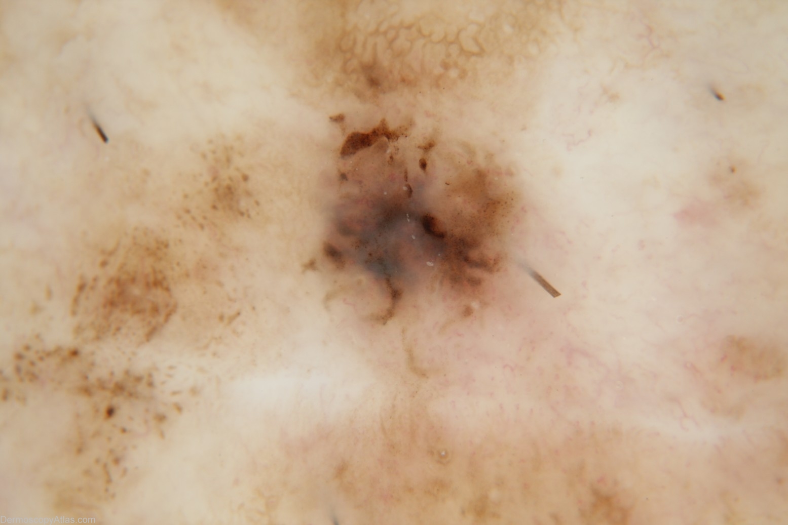
Description: Dermoscopy with Dermlite 2HR The eccentric focal brown dots and the polymorphous vessels are displayed more clearly in the polarised image.
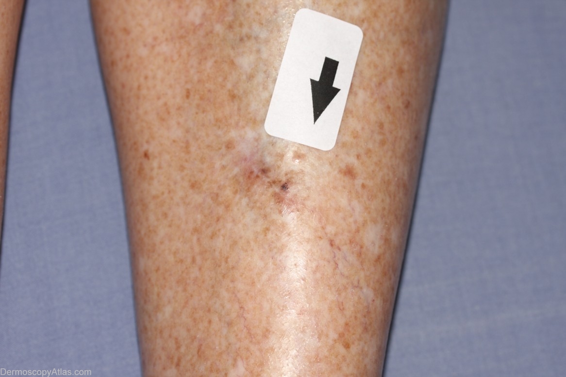
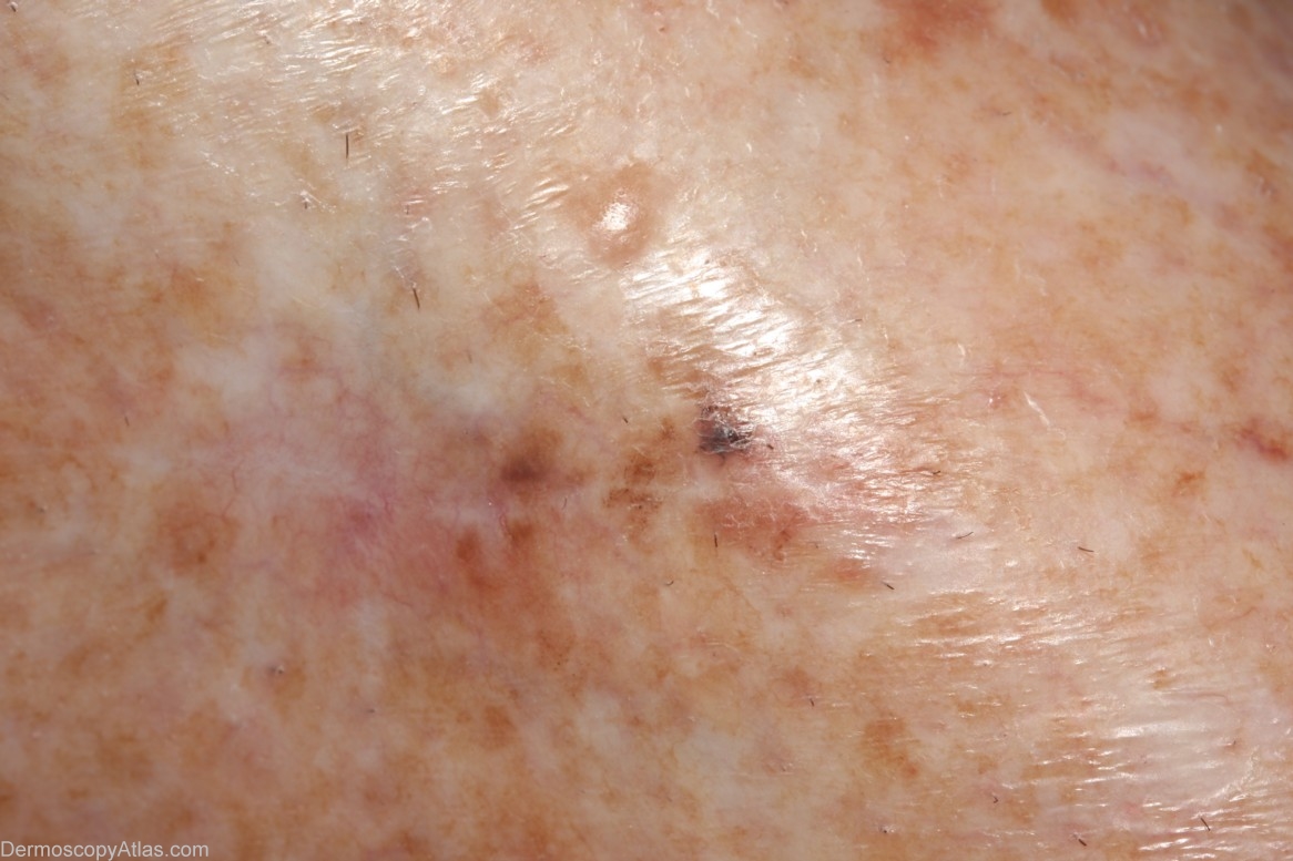
History: This 71 year old lady presented with this macule in continuity with a scar 4 years after excision of a level 4 lentigo maligna melanoma which had been excised with 1.5 mm clearance at the closest margin. The initial exision biopsy of the pigmented macule at the lower end of the scar revealed level 1 lentigo maligna melanoma involving the entirety of both margins. Two further attempts at surgical clearance resulted finally in a level 1 lentigo maligna melanoma with lateral clearance of greater than 10 mm.


