
Site: Scalp
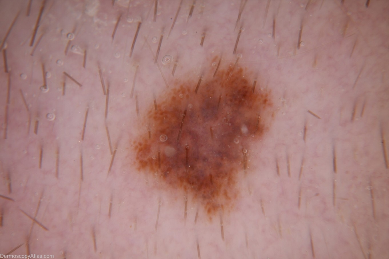
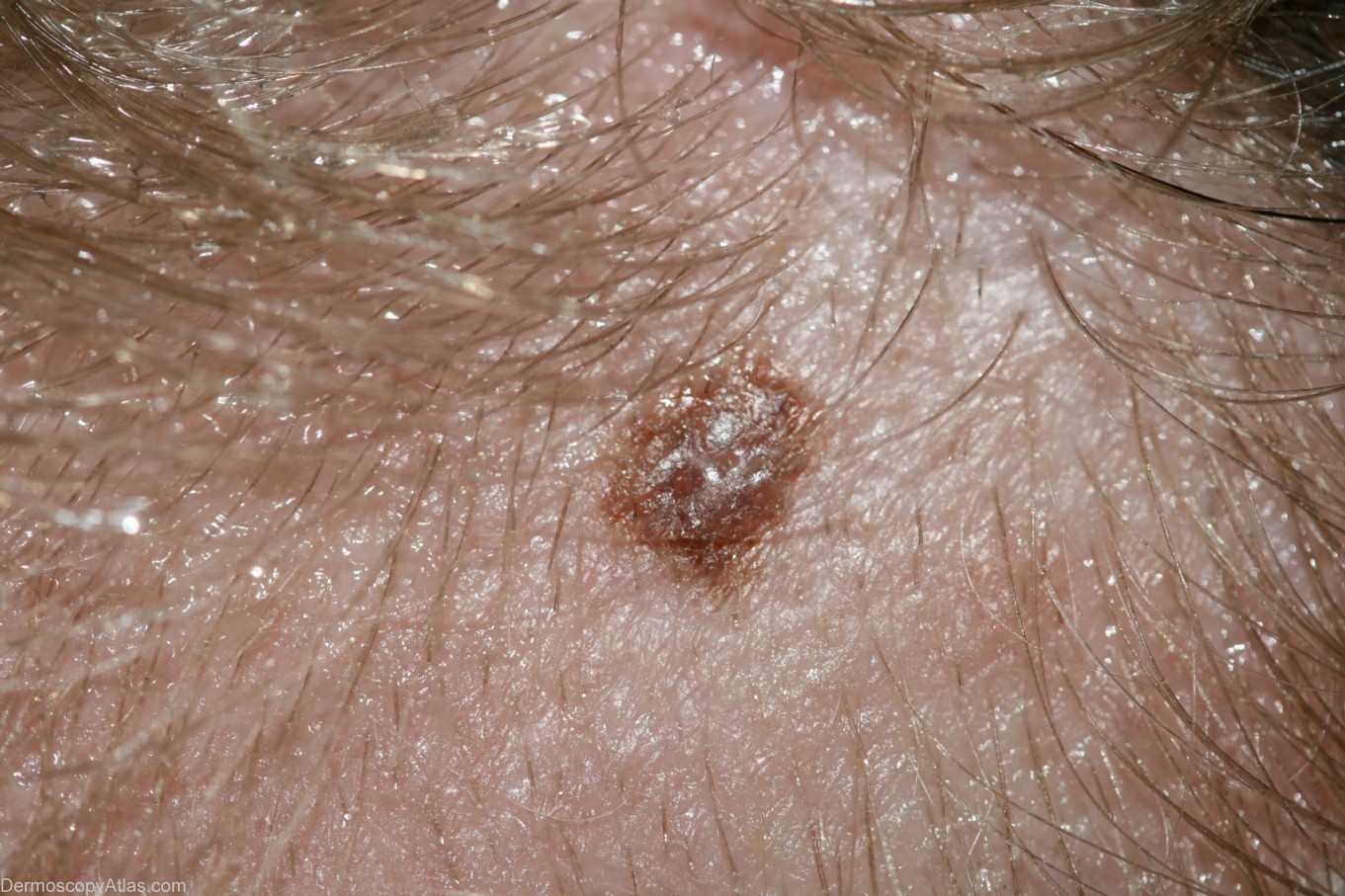
Diagnosis: Melanoma - Scalp location
Sex: F
Age: 20
Type: Dermlite Non Polarised
Submitted By: Cliff Rosendahl
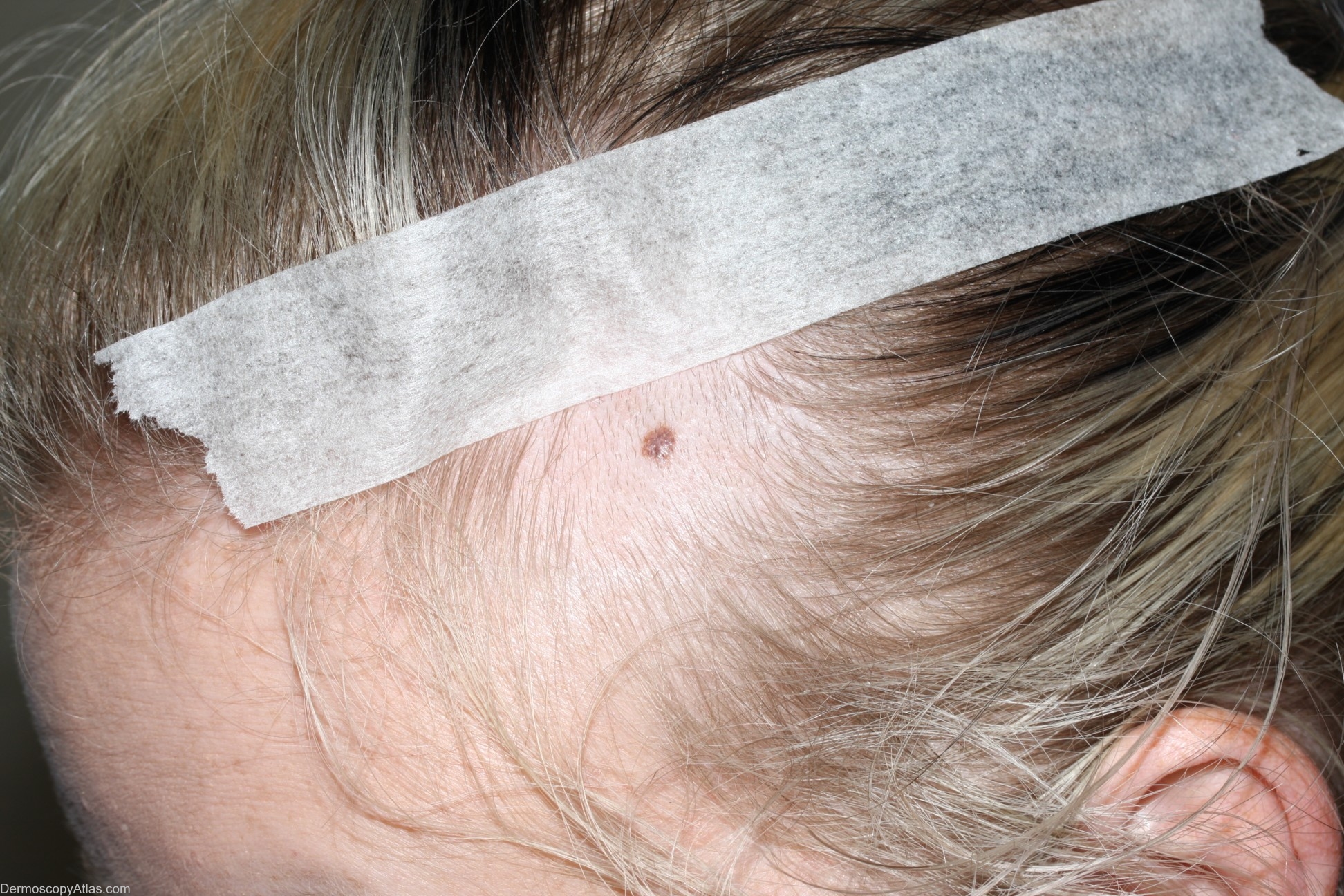
Description: Clinical image - hair has been clipped to facilitate dermoscopy. This site was completely hair covered and meaningful dermoscopy examination was not possible before hair-removal.
History:
This young lady had a borderline level 1 melanoma excised from her forearm at age 12. This scalp lesion was encountered at a routine skin examination and on specific questioning the patient believed that it had changed recently. Being on hair-protected scalp on a young person this was an unlikely site for a melanoma but with her past history and a history of change the lesion was excised. Histology was reported by David Weedon:-
"Sections show a level 2 (0.5mm thick) superficial spreading melanoma.
There is no ulceration or dermal mitoses. There is a mild lymphocytic
infiltrate with focal old regression only. The excisions appears
complete with lateral clearances of 1.6mm."
The histology slide has been sent to Harald Kittler. I will add his assessment here when he has reviewed the slide and he will also provide images of that slide which I will add to this posting.