
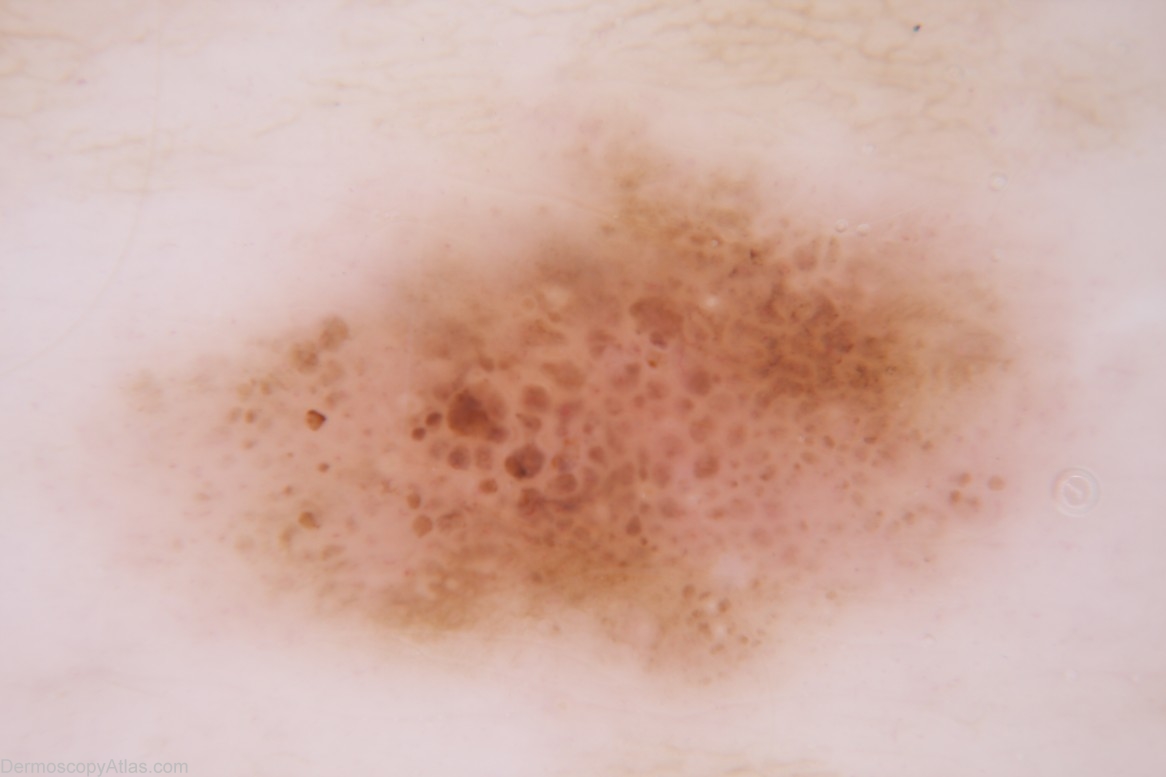
Site: Back
Diagnosis: Nevus dysplastic
Sex: M
Age: 45
Type: Dermlite Non Polarised
Submitted By: Cliff Rosendahl
Description: Dermoscopy image - Pattern - Brown clods, a small area with branched streaks and a small structureless area.Colours - Light brown, dark brown, pink and grey. Clues to melanoma - No convincing clues but the small structureless area with grey colour drew my attention
History: This 44 year old man has had two previous melanomas. These were a level 2 melanoma on the dorsum of the foot and a level 1 melanoma on the posterior surface of the ear (case 301 in this atlas). This lesion appeared to have changed and although it lacked definite clues to melanoma a specific confident benign diagnosis could not be made and it was thought to be too thick to safely monitor it. Histology revealed a naevus with "mild to moderate dysplasia".