
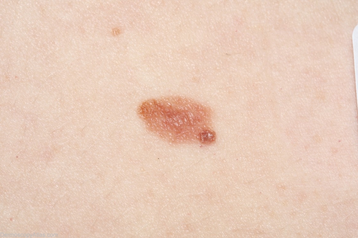
Site: Abdomen
Diagnosis: Nevus dysplastic
Sex: F
Age: 44
Type: Dermlite Non Polarised
Submitted By: Cliff Rosendahl
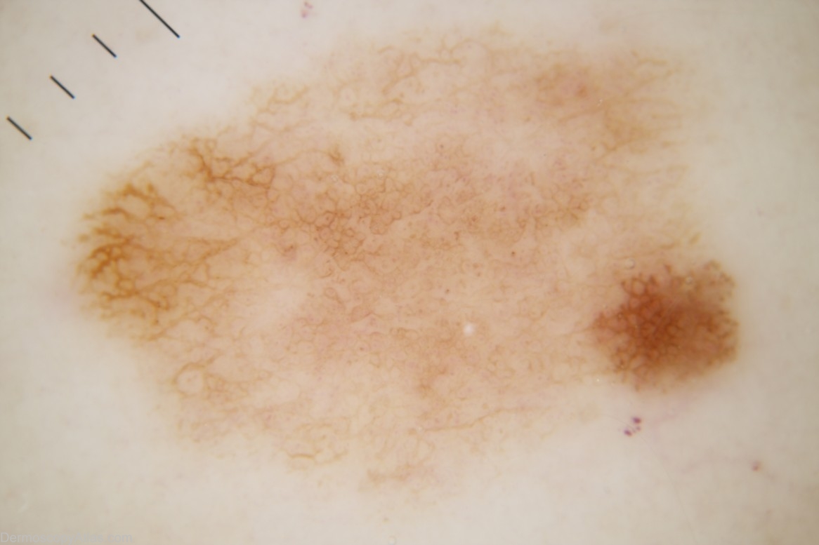
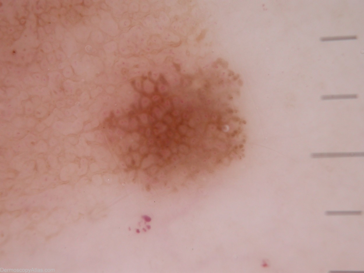
Description: There are peripheral brown dots in the hyperpigmented focus but as these can be found in any growing naevus they are not a clue to melanoma. There is one possible pseudopod which is not counted because it is equivocal.
History:
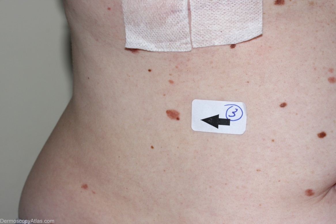
This 44 year old lady has had 3 previous melanomas. This lesion was noticed on her initial visit to me at which time I removed 2 melanomas (her 2nd and 3rd). This naevus was scheduled for removal a couple of weeks later. The histology report was as follows:-
" Sections show a bland intradermal naevus over the top of which has developed a multifocal junctional dysplastic naevus with mild to moderate atypia. Excision appears complete."
Harald Kittler's assessment prior to seeing the histology report was as follows:-
"Pattern: lines reticular
Colors: eccentric hyperpigmentation
Clues: peripheral brown dots are not a clue to melanoma, because they can be found in any growing lesion. There is a possible pseudopod at the SSE, but whenever I say possible and not striking, I do not count it.
Summary: DDX between Melanoma in situ and Clark's nevus cannot be resolved. With regard to the previous history of multiple melanomas, I would not hesitate to excise the lesion."